Chlorine »
PDB 2dct-2dxa »
2djf »
Chlorine in PDB 2djf: Crystal Structure of Human Dipeptidyl Peptidase I (Cathepsin C) in Complex with the Inhibitor Gly-Phe-CHN2
Enzymatic activity of Crystal Structure of Human Dipeptidyl Peptidase I (Cathepsin C) in Complex with the Inhibitor Gly-Phe-CHN2
All present enzymatic activity of Crystal Structure of Human Dipeptidyl Peptidase I (Cathepsin C) in Complex with the Inhibitor Gly-Phe-CHN2:
3.4.14.1;
3.4.14.1;
Protein crystallography data
The structure of Crystal Structure of Human Dipeptidyl Peptidase I (Cathepsin C) in Complex with the Inhibitor Gly-Phe-CHN2, PDB code: 2djf
was solved by
A.Molgaard,
J.Arnau,
C.Lauritzen,
S.Larsen,
G.Petersen,
J.Pedersen,
with X-Ray Crystallography technique. A brief refinement statistics is given in the table below:
| Resolution Low / High (Å) | 28.75 / 2.00 |
| Space group | I 2 2 2 |
| Cell size a, b, c (Å), α, β, γ (°) | 87.000, 89.030, 115.570, 90.00, 90.00, 90.00 |
| R / Rfree (%) | 16.1 / 20 |
Chlorine Binding Sites:
The binding sites of Chlorine atom in the Crystal Structure of Human Dipeptidyl Peptidase I (Cathepsin C) in Complex with the Inhibitor Gly-Phe-CHN2
(pdb code 2djf). This binding sites where shown within
5.0 Angstroms radius around Chlorine atom.
In total only one binding site of Chlorine was determined in the Crystal Structure of Human Dipeptidyl Peptidase I (Cathepsin C) in Complex with the Inhibitor Gly-Phe-CHN2, PDB code: 2djf:
In total only one binding site of Chlorine was determined in the Crystal Structure of Human Dipeptidyl Peptidase I (Cathepsin C) in Complex with the Inhibitor Gly-Phe-CHN2, PDB code: 2djf:
Chlorine binding site 1 out of 1 in 2djf
Go back to
Chlorine binding site 1 out
of 1 in the Crystal Structure of Human Dipeptidyl Peptidase I (Cathepsin C) in Complex with the Inhibitor Gly-Phe-CHN2
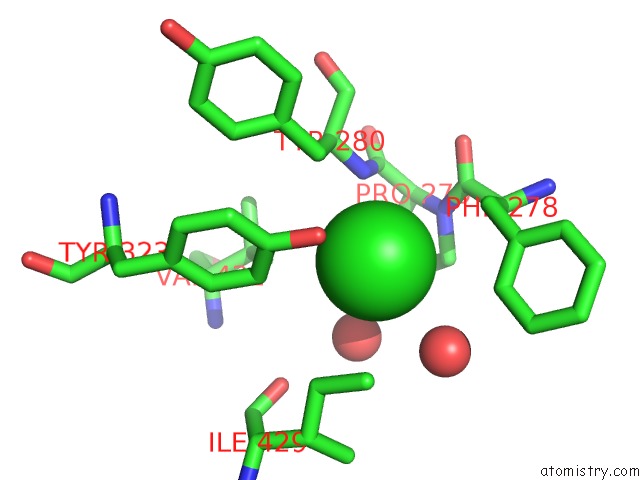
Mono view
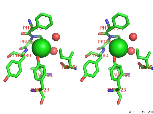
Stereo pair view
Mono view
Stereo pair view
A full contact list of Chlorine with other atoms in the Cl binding
site number 1 of Crystal Structure of Human Dipeptidyl Peptidase I (Cathepsin C) in Complex with the Inhibitor Gly-Phe-CHN2 within 5.0Å range:
|
Reference:
A.Molgaard,
J.Arnau,
C.Lauritzen,
S.Larsen,
G.Petersen,
J.Pedersen.
The Crystal Structure of Human Dipeptidyl Peptidase I (Cathepsin C) in Complex with the Inhibitor Gly-Phe-CHN2 Biochem.J. V. 401 645 2007.
ISSN: ISSN 0264-6021
PubMed: 17020538
DOI: 10.1042/BJ20061389
Page generated: Thu Jul 10 21:52:13 2025
ISSN: ISSN 0264-6021
PubMed: 17020538
DOI: 10.1042/BJ20061389
Last articles
Cl in 2WHVCl in 2WG3
Cl in 2WEK
Cl in 2WGJ
Cl in 2WET
Cl in 2WG2
Cl in 2WG0
Cl in 2WFZ
Cl in 2WFT
Cl in 2WDR