Chlorine »
PDB 2pgc-2px2 »
2puw »
Chlorine in PDB 2puw: The Crystal Structure of Isomerase Domain of Glucosamine-6-Phosphate Synthase From Candida Albicans
Enzymatic activity of The Crystal Structure of Isomerase Domain of Glucosamine-6-Phosphate Synthase From Candida Albicans
All present enzymatic activity of The Crystal Structure of Isomerase Domain of Glucosamine-6-Phosphate Synthase From Candida Albicans:
2.6.1.16;
2.6.1.16;
Protein crystallography data
The structure of The Crystal Structure of Isomerase Domain of Glucosamine-6-Phosphate Synthase From Candida Albicans, PDB code: 2puw
was solved by
J.Raczynska,
J.Olchowy,
S.Milewski,
W.Rypniewski,
with X-Ray Crystallography technique. A brief refinement statistics is given in the table below:
| Resolution Low / High (Å) | 19.55 / 3.15 |
| Space group | I 4 |
| Cell size a, b, c (Å), α, β, γ (°) | 148.864, 148.864, 102.878, 90.00, 90.00, 90.00 |
| R / Rfree (%) | 23 / 27.9 |
Chlorine Binding Sites:
The binding sites of Chlorine atom in the The Crystal Structure of Isomerase Domain of Glucosamine-6-Phosphate Synthase From Candida Albicans
(pdb code 2puw). This binding sites where shown within
5.0 Angstroms radius around Chlorine atom.
In total 2 binding sites of Chlorine where determined in the The Crystal Structure of Isomerase Domain of Glucosamine-6-Phosphate Synthase From Candida Albicans, PDB code: 2puw:
Jump to Chlorine binding site number: 1; 2;
In total 2 binding sites of Chlorine where determined in the The Crystal Structure of Isomerase Domain of Glucosamine-6-Phosphate Synthase From Candida Albicans, PDB code: 2puw:
Jump to Chlorine binding site number: 1; 2;
Chlorine binding site 1 out of 2 in 2puw
Go back to
Chlorine binding site 1 out
of 2 in the The Crystal Structure of Isomerase Domain of Glucosamine-6-Phosphate Synthase From Candida Albicans
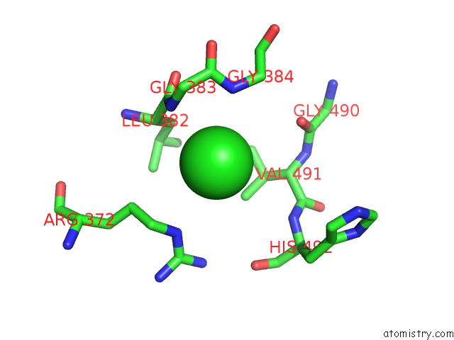
Mono view
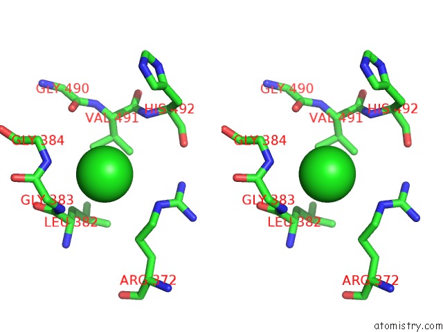
Stereo pair view
Mono view
Stereo pair view
A full contact list of Chlorine with other atoms in the Cl binding
site number 1 of The Crystal Structure of Isomerase Domain of Glucosamine-6-Phosphate Synthase From Candida Albicans within 5.0Å range:
|
Chlorine binding site 2 out of 2 in 2puw
Go back to
Chlorine binding site 2 out
of 2 in the The Crystal Structure of Isomerase Domain of Glucosamine-6-Phosphate Synthase From Candida Albicans
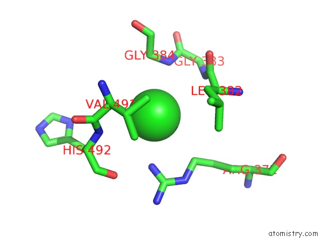
Mono view
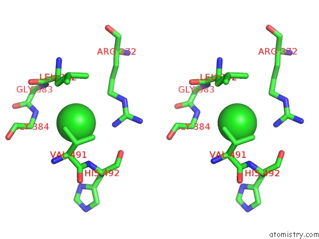
Stereo pair view
Mono view
Stereo pair view
A full contact list of Chlorine with other atoms in the Cl binding
site number 2 of The Crystal Structure of Isomerase Domain of Glucosamine-6-Phosphate Synthase From Candida Albicans within 5.0Å range:
|
Reference:
J.Raczynska,
J.Olchowy,
P.V.Konariev,
D.I.Svergun,
S.Milewski,
W.Rypniewski.
The Crystal and Solution Studies of Glucosamine-6-Phosphate Synthase From Candida Albicans J.Mol.Biol. V. 372 672 2007.
ISSN: ISSN 0022-2836
PubMed: 17681543
DOI: 10.1016/J.JMB.2007.07.002
Page generated: Thu Jul 10 23:55:46 2025
ISSN: ISSN 0022-2836
PubMed: 17681543
DOI: 10.1016/J.JMB.2007.07.002
Last articles
Cl in 3FAJCl in 3FAH
Cl in 3FAK
Cl in 3FA5
Cl in 3F9O
Cl in 3F95
Cl in 3F9P
Cl in 3FA0
Cl in 3F9N
Cl in 3F9L