Chlorine »
PDB 2wcz-2wjp »
2wjp »
Chlorine in PDB 2wjp: Crystal Structure of Murd Ligase in Complex with D-Glu Containing Rhodanine Inhibitor
Enzymatic activity of Crystal Structure of Murd Ligase in Complex with D-Glu Containing Rhodanine Inhibitor
All present enzymatic activity of Crystal Structure of Murd Ligase in Complex with D-Glu Containing Rhodanine Inhibitor:
6.3.2.9;
6.3.2.9;
Protein crystallography data
The structure of Crystal Structure of Murd Ligase in Complex with D-Glu Containing Rhodanine Inhibitor, PDB code: 2wjp
was solved by
T.Tomasic,
N.Zidar,
R.Sink,
A.Kovac,
V.Rupnik,
S.Turk,
C.Contreras-Martel,
A.Dessen,
D.Blanot,
M.Muller-Premru,
S.Gobec,
A.Zega,
L.Peterlin-Masic,
D.Kikelj,
with X-Ray Crystallography technique. A brief refinement statistics is given in the table below:
| Resolution Low / High (Å) | 46.63 / 1.60 |
| Space group | P 41 |
| Cell size a, b, c (Å), α, β, γ (°) | 65.917, 65.917, 136.063, 90.00, 90.00, 90.00 |
| R / Rfree (%) | 16.6 / 18.9 |
Chlorine Binding Sites:
The binding sites of Chlorine atom in the Crystal Structure of Murd Ligase in Complex with D-Glu Containing Rhodanine Inhibitor
(pdb code 2wjp). This binding sites where shown within
5.0 Angstroms radius around Chlorine atom.
In total 6 binding sites of Chlorine where determined in the Crystal Structure of Murd Ligase in Complex with D-Glu Containing Rhodanine Inhibitor, PDB code: 2wjp:
Jump to Chlorine binding site number: 1; 2; 3; 4; 5; 6;
In total 6 binding sites of Chlorine where determined in the Crystal Structure of Murd Ligase in Complex with D-Glu Containing Rhodanine Inhibitor, PDB code: 2wjp:
Jump to Chlorine binding site number: 1; 2; 3; 4; 5; 6;
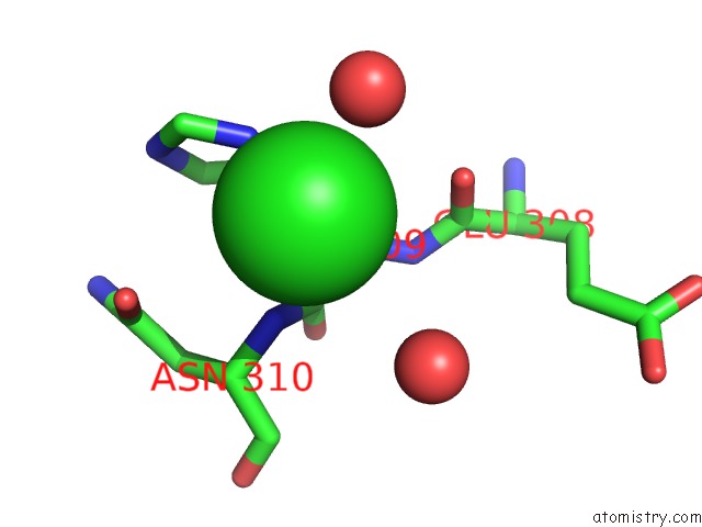
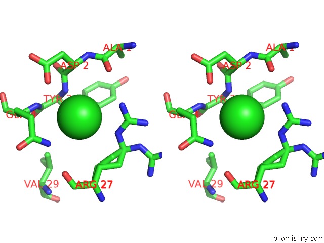
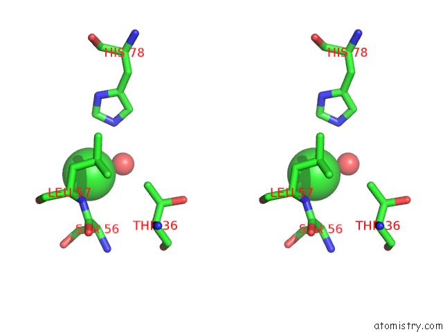
Chlorine binding site 1 out of 6 in 2wjp
Go back to
Chlorine binding site 1 out
of 6 in the Crystal Structure of Murd Ligase in Complex with D-Glu Containing Rhodanine Inhibitor
Mono view

Stereo pair view
Mono view
Stereo pair view
A full contact list of Chlorine with other atoms in the Cl binding
site number 1 of Crystal Structure of Murd Ligase in Complex with D-Glu Containing Rhodanine Inhibitor within 5.0Å range:
|
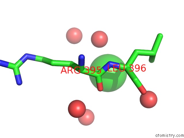
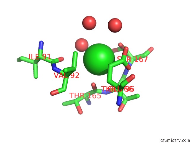
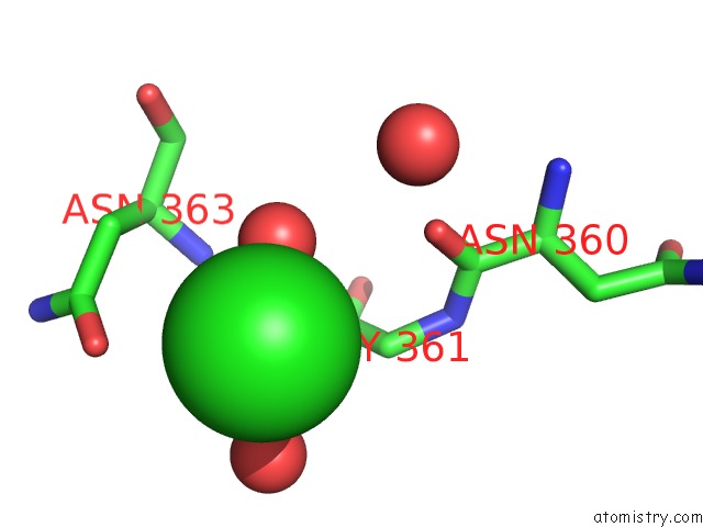
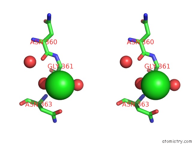
Chlorine binding site 2 out of 6 in 2wjp
Go back to
Chlorine binding site 2 out
of 6 in the Crystal Structure of Murd Ligase in Complex with D-Glu Containing Rhodanine Inhibitor
Mono view
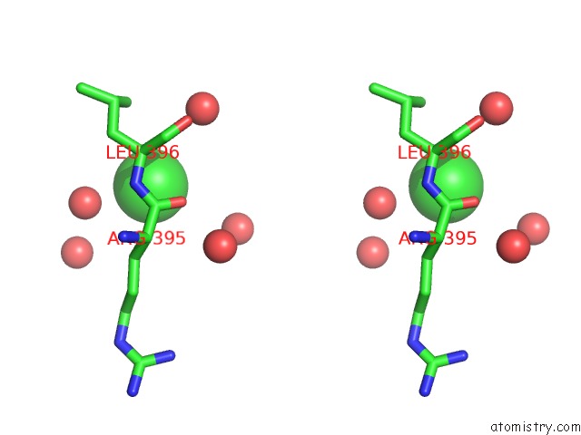
Stereo pair view
Mono view
Stereo pair view
A full contact list of Chlorine with other atoms in the Cl binding
site number 2 of Crystal Structure of Murd Ligase in Complex with D-Glu Containing Rhodanine Inhibitor within 5.0Å range:
|
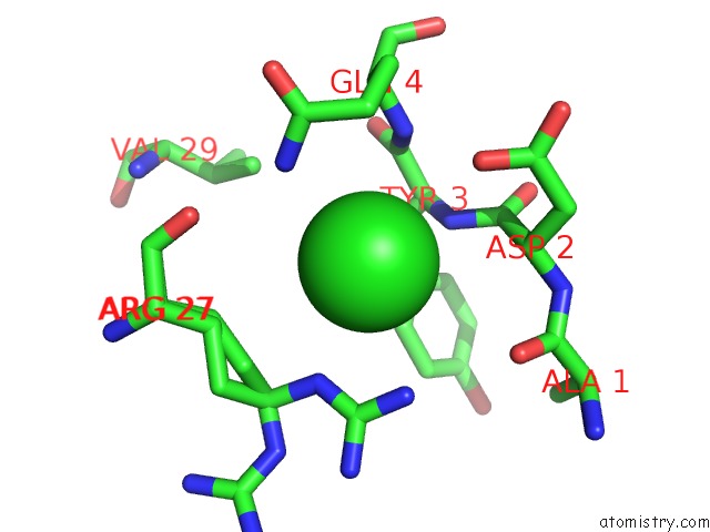
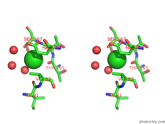
Chlorine binding site 3 out of 6 in 2wjp
Go back to
Chlorine binding site 3 out
of 6 in the Crystal Structure of Murd Ligase in Complex with D-Glu Containing Rhodanine Inhibitor
Mono view
Stereo pair view
Mono view
Stereo pair view
A full contact list of Chlorine with other atoms in the Cl binding
site number 3 of Crystal Structure of Murd Ligase in Complex with D-Glu Containing Rhodanine Inhibitor within 5.0Å range:
|
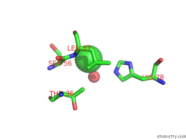
Chlorine binding site 4 out of 6 in 2wjp
Go back to
Chlorine binding site 4 out
of 6 in the Crystal Structure of Murd Ligase in Complex with D-Glu Containing Rhodanine Inhibitor
Mono view
Stereo pair view
Mono view
Stereo pair view
A full contact list of Chlorine with other atoms in the Cl binding
site number 4 of Crystal Structure of Murd Ligase in Complex with D-Glu Containing Rhodanine Inhibitor within 5.0Å range:
|
Chlorine binding site 5 out of 6 in 2wjp
Go back to
Chlorine binding site 5 out
of 6 in the Crystal Structure of Murd Ligase in Complex with D-Glu Containing Rhodanine Inhibitor
Mono view
Stereo pair view
Mono view
Stereo pair view
A full contact list of Chlorine with other atoms in the Cl binding
site number 5 of Crystal Structure of Murd Ligase in Complex with D-Glu Containing Rhodanine Inhibitor within 5.0Å range:
|
Chlorine binding site 6 out of 6 in 2wjp
Go back to
Chlorine binding site 6 out
of 6 in the Crystal Structure of Murd Ligase in Complex with D-Glu Containing Rhodanine Inhibitor
Mono view
Stereo pair view
Mono view
Stereo pair view
A full contact list of Chlorine with other atoms in the Cl binding
site number 6 of Crystal Structure of Murd Ligase in Complex with D-Glu Containing Rhodanine Inhibitor within 5.0Å range:
|
Reference:
N.Zidar,
T.Tomasic,
R.Sink,
V.Rupnik,
A.Kovac,
S.Turk,
D.Patin,
D.Blanot,
C.Contreras Martel,
A.Dessen,
M.Muller Premru,
A.Zega,
S.Gobec,
L.Peterlin Masic,
D.Kikelj.
Discovery of Novel 5-Benzylidenerhodanine and 5- Benzylidenethiazolidine-2,4-Dione Inhibitors of Murd Ligase. J.Med.Chem. V. 53 6584 2010.
ISSN: ISSN 0022-2623
PubMed: 20804196
DOI: 10.1021/JM100285G
Page generated: Fri Jul 11 01:27:36 2025
ISSN: ISSN 0022-2623
PubMed: 20804196
DOI: 10.1021/JM100285G
Last articles
Cl in 5VTKCl in 5VSV
Cl in 5VR0
Cl in 5VTI
Cl in 5VTD
Cl in 5VT7
Cl in 5VSK
Cl in 5VSB
Cl in 5VS7
Cl in 5VPB