Chlorine »
PDB 2y5f-2yc5 »
2y5t »
Chlorine in PDB 2y5t: Crystal Structure of the Pathogenic Autoantibody CIIC1 in Complex with the Triple-Helical C1 Peptide
Protein crystallography data
The structure of Crystal Structure of the Pathogenic Autoantibody CIIC1 in Complex with the Triple-Helical C1 Peptide, PDB code: 2y5t
was solved by
D.Dobritzsch,
I.Lindh,
N.Schneider,
H.Uysal,
K.S.Nandakumar,
H.Burkhardt,
G.Schneider,
R.Holmdahl,
with X-Ray Crystallography technique. A brief refinement statistics is given in the table below:
| Resolution Low / High (Å) | 43.85 / 2.20 |
| Space group | P 31 2 1 |
| Cell size a, b, c (Å), α, β, γ (°) | 81.050, 81.050, 168.450, 90.00, 90.00, 120.00 |
| R / Rfree (%) | 18.8 / 24.5 |
Chlorine Binding Sites:
The binding sites of Chlorine atom in the Crystal Structure of the Pathogenic Autoantibody CIIC1 in Complex with the Triple-Helical C1 Peptide
(pdb code 2y5t). This binding sites where shown within
5.0 Angstroms radius around Chlorine atom.
In total only one binding site of Chlorine was determined in the Crystal Structure of the Pathogenic Autoantibody CIIC1 in Complex with the Triple-Helical C1 Peptide, PDB code: 2y5t:
In total only one binding site of Chlorine was determined in the Crystal Structure of the Pathogenic Autoantibody CIIC1 in Complex with the Triple-Helical C1 Peptide, PDB code: 2y5t:
Chlorine binding site 1 out of 1 in 2y5t
Go back to
Chlorine binding site 1 out
of 1 in the Crystal Structure of the Pathogenic Autoantibody CIIC1 in Complex with the Triple-Helical C1 Peptide
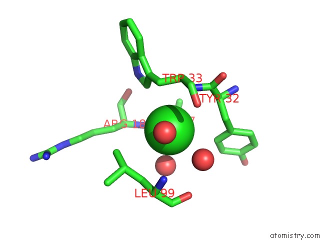
Mono view
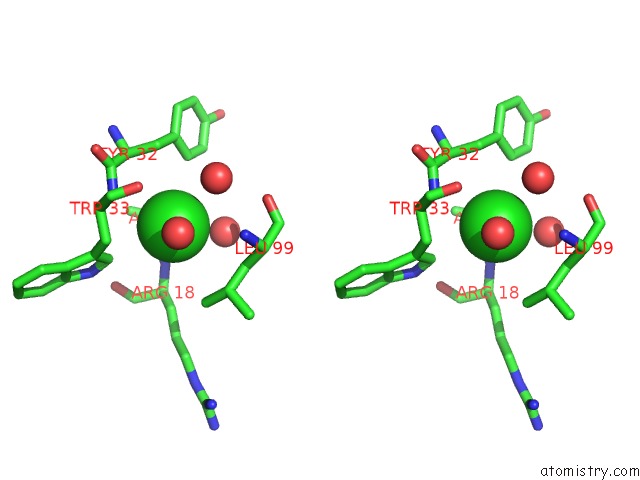
Stereo pair view
Mono view
Stereo pair view
A full contact list of Chlorine with other atoms in the Cl binding
site number 1 of Crystal Structure of the Pathogenic Autoantibody CIIC1 in Complex with the Triple-Helical C1 Peptide within 5.0Å range:
|
Reference:
D.Dobritzsch,
I.Lindh,
H.Uysal,
K.S.Nandakumar,
H.Burkhardt,
G.Schneider,
R.Holmdahl.
Crystal Structure of An Arthritogenic Anticollagen Immune Complex. Arthritis Rheum. V. 63 3740 2011.
ISSN: ISSN 0004-3591
PubMed: 22127694
DOI: 10.1002/ART.30611
Page generated: Fri Jul 11 02:30:54 2025
ISSN: ISSN 0004-3591
PubMed: 22127694
DOI: 10.1002/ART.30611
Last articles
Cl in 3OCBCl in 3OCA
Cl in 3OC2
Cl in 3OAG
Cl in 3OC1
Cl in 3OBJ
Cl in 3OAD
Cl in 3OB4
Cl in 3OAO
Cl in 3OAF