Chlorine »
PDB 2z0g-2zgd »
2ze5 »
Chlorine in PDB 2ze5: Crystal Structure of Adenosine Phosphate-Isopentenyltransferase
Enzymatic activity of Crystal Structure of Adenosine Phosphate-Isopentenyltransferase
All present enzymatic activity of Crystal Structure of Adenosine Phosphate-Isopentenyltransferase:
2.5.1.27;
2.5.1.27;
Protein crystallography data
The structure of Crystal Structure of Adenosine Phosphate-Isopentenyltransferase, PDB code: 2ze5
was solved by
H.Sakakibara,
with X-Ray Crystallography technique. A brief refinement statistics is given in the table below:
| Resolution Low / High (Å) | 68.20 / 2.31 |
| Space group | P 42 21 2 |
| Cell size a, b, c (Å), α, β, γ (°) | 96.392, 96.392, 65.197, 90.00, 90.00, 90.00 |
| R / Rfree (%) | 20.1 / 25.4 |
Chlorine Binding Sites:
The binding sites of Chlorine atom in the Crystal Structure of Adenosine Phosphate-Isopentenyltransferase
(pdb code 2ze5). This binding sites where shown within
5.0 Angstroms radius around Chlorine atom.
In total only one binding site of Chlorine was determined in the Crystal Structure of Adenosine Phosphate-Isopentenyltransferase, PDB code: 2ze5:
In total only one binding site of Chlorine was determined in the Crystal Structure of Adenosine Phosphate-Isopentenyltransferase, PDB code: 2ze5:
Chlorine binding site 1 out of 1 in 2ze5
Go back to
Chlorine binding site 1 out
of 1 in the Crystal Structure of Adenosine Phosphate-Isopentenyltransferase
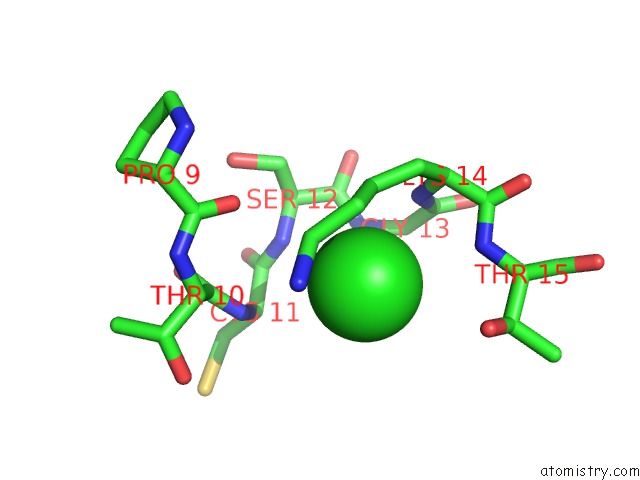
Mono view
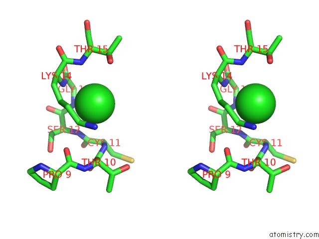
Stereo pair view
Mono view
Stereo pair view
A full contact list of Chlorine with other atoms in the Cl binding
site number 1 of Crystal Structure of Adenosine Phosphate-Isopentenyltransferase within 5.0Å range:
|
Reference:
H.Sugawara,
N.Ueda,
M.Kojima,
N.Makita,
T.Yamaya,
H.Sakakibara.
Structural Insight Into the Reaction Mechanism and Evolution of Cytokinin Biosynthesis. Proc.Natl.Acad.Sci.Usa V. 105 2734 2008.
ISSN: ISSN 0027-8424
PubMed: 18258747
DOI: 10.1073/PNAS.0707374105
Page generated: Fri Jul 11 02:54:23 2025
ISSN: ISSN 0027-8424
PubMed: 18258747
DOI: 10.1073/PNAS.0707374105
Last articles
Cl in 3OLDCl in 3OKT
Cl in 3OKI
Cl in 3OKH
Cl in 3OJT
Cl in 3OID
Cl in 3OJP
Cl in 3OK0
Cl in 3OJN
Cl in 3OJK