Chlorine »
PDB 3b0x-3bft »
3bcb »
Chlorine in PDB 3bcb: Crystal Structure of Mouse Selenocysteine Synthase, Sodium Phosphate Soak
Protein crystallography data
The structure of Crystal Structure of Mouse Selenocysteine Synthase, Sodium Phosphate Soak, PDB code: 3bcb
was solved by
O.M.Ganichkin,
M.C.Wahl,
with X-Ray Crystallography technique. A brief refinement statistics is given in the table below:
| Resolution Low / High (Å) | 20.00 / 1.85 |
| Space group | I 2 2 2 |
| Cell size a, b, c (Å), α, β, γ (°) | 59.231, 138.974, 141.821, 90.00, 90.00, 90.00 |
| R / Rfree (%) | 16.6 / 20.3 |
Chlorine Binding Sites:
The binding sites of Chlorine atom in the Crystal Structure of Mouse Selenocysteine Synthase, Sodium Phosphate Soak
(pdb code 3bcb). This binding sites where shown within
5.0 Angstroms radius around Chlorine atom.
In total only one binding site of Chlorine was determined in the Crystal Structure of Mouse Selenocysteine Synthase, Sodium Phosphate Soak, PDB code: 3bcb:
In total only one binding site of Chlorine was determined in the Crystal Structure of Mouse Selenocysteine Synthase, Sodium Phosphate Soak, PDB code: 3bcb:
Chlorine binding site 1 out of 1 in 3bcb
Go back to
Chlorine binding site 1 out
of 1 in the Crystal Structure of Mouse Selenocysteine Synthase, Sodium Phosphate Soak
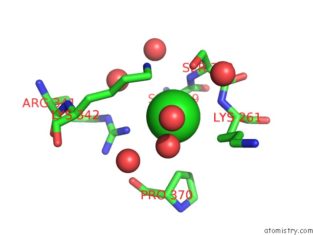
Mono view
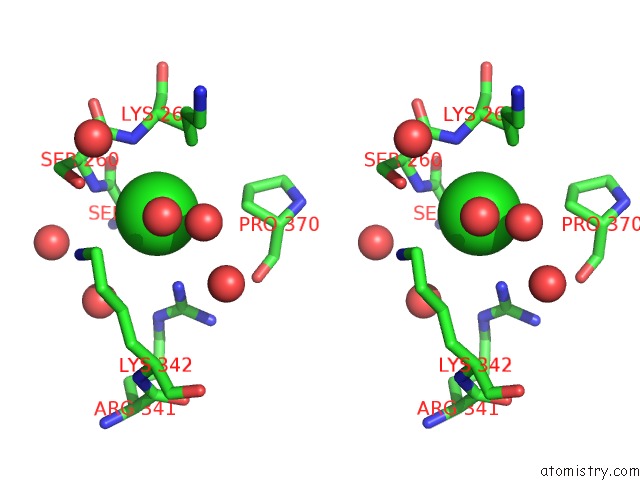
Stereo pair view
Mono view
Stereo pair view
A full contact list of Chlorine with other atoms in the Cl binding
site number 1 of Crystal Structure of Mouse Selenocysteine Synthase, Sodium Phosphate Soak within 5.0Å range:
|
Reference:
O.M.Ganichkin,
X.M.Xu,
B.A.Carlson,
H.Mix,
D.L.Hatfield,
V.N.Gladyshev,
M.C.Wahl.
Structure and Catalytic Mechanism of Eukaryotic Selenocysteine Synthase. J.Biol.Chem. V. 283 5849 2008.
ISSN: ISSN 0021-9258
PubMed: 18093968
DOI: 10.1074/JBC.M709342200
Page generated: Fri Jul 11 03:25:09 2025
ISSN: ISSN 0021-9258
PubMed: 18093968
DOI: 10.1074/JBC.M709342200
Last articles
Cl in 3OFWCl in 3OG3
Cl in 3OAZ
Cl in 3OEX
Cl in 3OFM
Cl in 3OEU
Cl in 3OBK
Cl in 3ODG
Cl in 3OCT
Cl in 3OCL