Chlorine »
PDB 3dna-3dzc »
3dxb »
Chlorine in PDB 3dxb: Structure of the Uhm Domain of PUF60 Fused to Thioredoxin
Protein crystallography data
The structure of Structure of the Uhm Domain of PUF60 Fused to Thioredoxin, PDB code: 3dxb
was solved by
L.Corsini,
M.Hothorn,
K.Scheffzek,
G.Stier,
M.Sattler,
with X-Ray Crystallography technique. A brief refinement statistics is given in the table below:
| Resolution Low / High (Å) | 49.75 / 2.20 |
| Space group | P 21 21 21 |
| Cell size a, b, c (Å), α, β, γ (°) | 75.120, 89.430, 299.390, 90.00, 90.00, 90.00 |
| R / Rfree (%) | 21.1 / 27.1 |
Chlorine Binding Sites:
The binding sites of Chlorine atom in the Structure of the Uhm Domain of PUF60 Fused to Thioredoxin
(pdb code 3dxb). This binding sites where shown within
5.0 Angstroms radius around Chlorine atom.
In total 4 binding sites of Chlorine where determined in the Structure of the Uhm Domain of PUF60 Fused to Thioredoxin, PDB code: 3dxb:
Jump to Chlorine binding site number: 1; 2; 3; 4;
In total 4 binding sites of Chlorine where determined in the Structure of the Uhm Domain of PUF60 Fused to Thioredoxin, PDB code: 3dxb:
Jump to Chlorine binding site number: 1; 2; 3; 4;
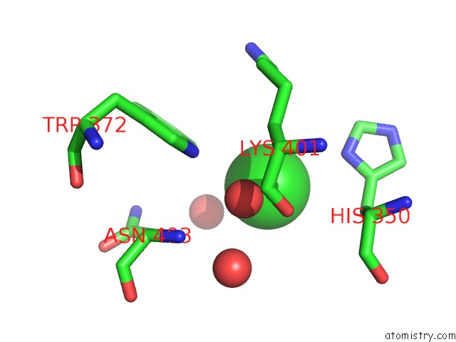
Chlorine binding site 1 out of 4 in 3dxb
Go back to
Chlorine binding site 1 out
of 4 in the Structure of the Uhm Domain of PUF60 Fused to Thioredoxin
Mono view

Stereo pair view
Mono view
Stereo pair view
A full contact list of Chlorine with other atoms in the Cl binding
site number 1 of Structure of the Uhm Domain of PUF60 Fused to Thioredoxin within 5.0Å range:
|
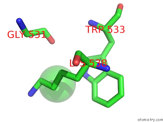
Chlorine binding site 2 out of 4 in 3dxb
Go back to
Chlorine binding site 2 out
of 4 in the Structure of the Uhm Domain of PUF60 Fused to Thioredoxin
Mono view

Stereo pair view
Mono view
Stereo pair view
A full contact list of Chlorine with other atoms in the Cl binding
site number 2 of Structure of the Uhm Domain of PUF60 Fused to Thioredoxin within 5.0Å range:
|
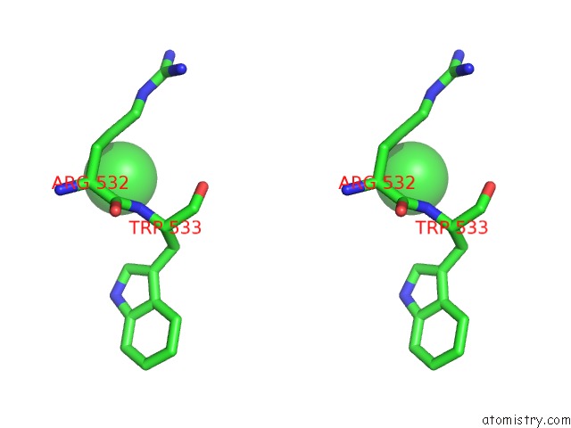
Chlorine binding site 3 out of 4 in 3dxb
Go back to
Chlorine binding site 3 out
of 4 in the Structure of the Uhm Domain of PUF60 Fused to Thioredoxin

Mono view
Stereo pair view
Mono view
Stereo pair view
A full contact list of Chlorine with other atoms in the Cl binding
site number 3 of Structure of the Uhm Domain of PUF60 Fused to Thioredoxin within 5.0Å range:
|
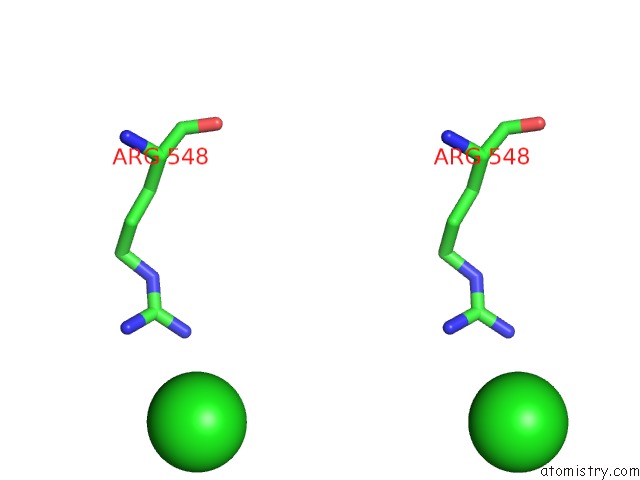
Chlorine binding site 4 out of 4 in 3dxb
Go back to
Chlorine binding site 4 out
of 4 in the Structure of the Uhm Domain of PUF60 Fused to Thioredoxin

Mono view
Stereo pair view
Mono view
Stereo pair view
A full contact list of Chlorine with other atoms in the Cl binding
site number 4 of Structure of the Uhm Domain of PUF60 Fused to Thioredoxin within 5.0Å range:
|
Reference:
L.Corsini,
M.Hothorn,
G.Stier,
V.Rybin,
K.Scheffzek,
T.J.Gibson,
M.Sattler.
Dimerization and Protein Binding Specificity of the U2AF Homology Motif of the Splicing Factor PUF60. J.Biol.Chem. V. 284 630 2009.
ISSN: ISSN 0021-9258
PubMed: 18974054
DOI: 10.1074/JBC.M805395200
Page generated: Fri Jul 11 04:39:25 2025
ISSN: ISSN 0021-9258
PubMed: 18974054
DOI: 10.1074/JBC.M805395200
Last articles
Fe in 2YXOFe in 2YRS
Fe in 2YXC
Fe in 2YNM
Fe in 2YVJ
Fe in 2YP1
Fe in 2YU2
Fe in 2YU1
Fe in 2YQB
Fe in 2YOO