Chlorine »
PDB 3ilj-3isv »
3isq »
Chlorine in PDB 3isq: Crystal Structure of Human 4-Hydroxyphenylpyruvate Dioxygenase
Enzymatic activity of Crystal Structure of Human 4-Hydroxyphenylpyruvate Dioxygenase
All present enzymatic activity of Crystal Structure of Human 4-Hydroxyphenylpyruvate Dioxygenase:
1.13.11.27;
1.13.11.27;
Protein crystallography data
The structure of Crystal Structure of Human 4-Hydroxyphenylpyruvate Dioxygenase, PDB code: 3isq
was solved by
E.S.Pilka,
N.Shafqat,
R.Cocking,
J.E.Bray,
T.Krojer,
A.C.W.Pike,
F.Vondelft,
W.W.Yue,
C.H.Arrowsmith,
J.Weigelt,
A.Edwards,
C.Bountra,
U.Oppermann,
K.L.Kavanagh,
Structural Genomics Consortium (Sgc),
with X-Ray Crystallography technique. A brief refinement statistics is given in the table below:
| Resolution Low / High (Å) | 49.60 / 1.75 |
| Space group | P 31 2 1 |
| Cell size a, b, c (Å), α, β, γ (°) | 99.206, 99.206, 87.726, 90.00, 90.00, 120.00 |
| R / Rfree (%) | 14.4 / 17.4 |
Other elements in 3isq:
The structure of Crystal Structure of Human 4-Hydroxyphenylpyruvate Dioxygenase also contains other interesting chemical elements:
| Cobalt | (Co) | 1 atom |
| Sodium | (Na) | 1 atom |
Chlorine Binding Sites:
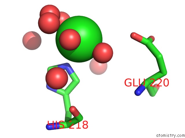
The binding sites of Chlorine atom in the Crystal Structure of Human 4-Hydroxyphenylpyruvate Dioxygenase
(pdb code 3isq). This binding sites where shown within
5.0 Angstroms radius around Chlorine atom.
In total only one binding site of Chlorine was determined in the Crystal Structure of Human 4-Hydroxyphenylpyruvate Dioxygenase, PDB code: 3isq:
In total only one binding site of Chlorine was determined in the Crystal Structure of Human 4-Hydroxyphenylpyruvate Dioxygenase, PDB code: 3isq:
Chlorine binding site 1 out of 1 in 3isq
Go back to
Chlorine binding site 1 out
of 1 in the Crystal Structure of Human 4-Hydroxyphenylpyruvate Dioxygenase
Mono view
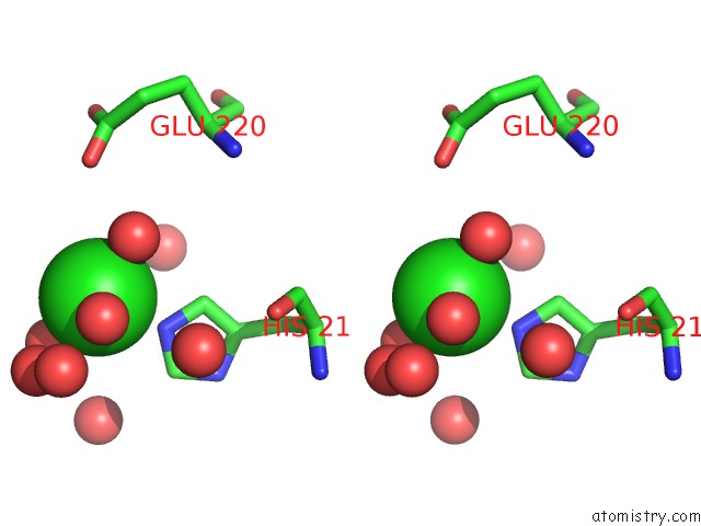
Stereo pair view
Mono view
Stereo pair view
A full contact list of Chlorine with other atoms in the Cl binding
site number 1 of Crystal Structure of Human 4-Hydroxyphenylpyruvate Dioxygenase within 5.0Å range:
|
Reference:
E.S.Pilka,
N.Shafqat,
R.Cocking,
J.E.Bray,
T.Krojer,
A.C.W.Pike,
F.Von Delft,
W.W.Yue,
C.H.Arrowsmith,
J.Weigelt,
A.Edwards,
C.Bountra,
U.Oppermann,
K.L.Kavanagh.
Crystal Structure of Human 4-Hydroxyphenylpyruvate Dioxygenase To Be Published.
Page generated: Sat Jul 20 21:52:05 2024
Last articles
Cl in 2W1PCl in 2W0U
Cl in 2VYX
Cl in 2W0R
Cl in 2W0D
Cl in 2VX3
Cl in 2VZ5
Cl in 2VYO
Cl in 2VYA
Cl in 2VY3