Chlorine »
PDB 3qp5-3qvm »
3qr8 »
Chlorine in PDB 3qr8: Crystal Structure of the Bacteriophage P2 Membrane-Piercing Protein Gpv
Protein crystallography data
The structure of Crystal Structure of the Bacteriophage P2 Membrane-Piercing Protein Gpv, PDB code: 3qr8
was solved by
C.Browning,
M.Shneider,
P.G.Leiman,
with X-Ray Crystallography technique. A brief refinement statistics is given in the table below:
| Resolution Low / High (Å) | 66.09 / 2.03 |
| Space group | P 3 2 1 |
| Cell size a, b, c (Å), α, β, γ (°) | 68.539, 68.539, 132.180, 90.00, 90.00, 120.00 |
| R / Rfree (%) | 26 / 30.5 |
Chlorine Binding Sites:
The binding sites of Chlorine atom in the Crystal Structure of the Bacteriophage P2 Membrane-Piercing Protein Gpv
(pdb code 3qr8). This binding sites where shown within
5.0 Angstroms radius around Chlorine atom.
In total only one binding site of Chlorine was determined in the Crystal Structure of the Bacteriophage P2 Membrane-Piercing Protein Gpv, PDB code: 3qr8:
In total only one binding site of Chlorine was determined in the Crystal Structure of the Bacteriophage P2 Membrane-Piercing Protein Gpv, PDB code: 3qr8:
Chlorine binding site 1 out of 1 in 3qr8
Go back to
Chlorine binding site 1 out
of 1 in the Crystal Structure of the Bacteriophage P2 Membrane-Piercing Protein Gpv
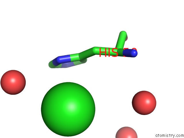
Mono view
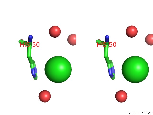
Stereo pair view
Mono view
Stereo pair view
A full contact list of Chlorine with other atoms in the Cl binding
site number 1 of Crystal Structure of the Bacteriophage P2 Membrane-Piercing Protein Gpv within 5.0Å range:
|
Reference:
C.Browning,
M.M.Shneider,
V.D.Bowman,
D.Schwarzer,
P.G.Leiman.
Phage Pierces the Host Cell Membrane with the Iron-Loaded Spike. Structure V. 20 326 2012.
ISSN: ISSN 0969-2126
PubMed: 22325780
DOI: 10.1016/J.STR.2011.12.009
Page generated: Fri Jul 11 09:31:19 2025
ISSN: ISSN 0969-2126
PubMed: 22325780
DOI: 10.1016/J.STR.2011.12.009
Last articles
Cl in 4HDPCl in 4HE0
Cl in 4HDT
Cl in 4HDC
Cl in 4HDK
Cl in 4HDF
Cl in 4HC6
Cl in 4HDB
Cl in 4HCX
Cl in 4HCD