Chlorine »
PDB 4e0j-4e9v »
4e1s »
Chlorine in PDB 4e1s: X-Ray Crystal Structure of the Transmembrane Beta-Domain From Intimin From Ehec Strain O157:H7
Protein crystallography data
The structure of X-Ray Crystal Structure of the Transmembrane Beta-Domain From Intimin From Ehec Strain O157:H7, PDB code: 4e1s
was solved by
J.W.Fairman,
N.Dautin,
D.Wojtowicz,
L.Wei,
N.Noinaj,
T.J.Barnard,
E.Udho,
A.Finkelstein,
T.M.Przytycka,
V.Cherezov,
S.K.Buchanan,
with X-Ray Crystallography technique. A brief refinement statistics is given in the table below:
| Resolution Low / High (Å) | 27.90 / 1.85 |
| Space group | C 2 2 21 |
| Cell size a, b, c (Å), α, β, γ (°) | 116.565, 120.248, 39.093, 90.00, 90.00, 90.00 |
| R / Rfree (%) | 17.5 / 23.3 |
Chlorine Binding Sites:
The binding sites of Chlorine atom in the X-Ray Crystal Structure of the Transmembrane Beta-Domain From Intimin From Ehec Strain O157:H7
(pdb code 4e1s). This binding sites where shown within
5.0 Angstroms radius around Chlorine atom.
In total only one binding site of Chlorine was determined in the X-Ray Crystal Structure of the Transmembrane Beta-Domain From Intimin From Ehec Strain O157:H7, PDB code: 4e1s:
In total only one binding site of Chlorine was determined in the X-Ray Crystal Structure of the Transmembrane Beta-Domain From Intimin From Ehec Strain O157:H7, PDB code: 4e1s:
Chlorine binding site 1 out of 1 in 4e1s
Go back to
Chlorine binding site 1 out
of 1 in the X-Ray Crystal Structure of the Transmembrane Beta-Domain From Intimin From Ehec Strain O157:H7
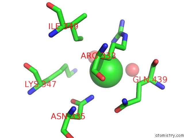
Mono view
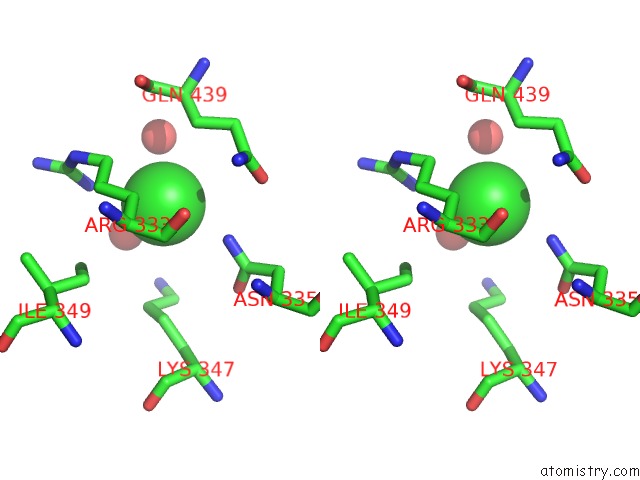
Stereo pair view
Mono view
Stereo pair view
A full contact list of Chlorine with other atoms in the Cl binding
site number 1 of X-Ray Crystal Structure of the Transmembrane Beta-Domain From Intimin From Ehec Strain O157:H7 within 5.0Å range:
|
Reference:
J.W.Fairman,
N.Dautin,
D.Wojtowicz,
W.Liu,
N.Noinaj,
T.J.Barnard,
E.Udho,
T.M.Przytycka,
V.Cherezov,
S.K.Buchanan.
Crystal Structures of the Outer Membrane Domain of Intimin and Invasin From Enterohemorrhagic E. Coli and Enteropathogenic Y. Pseudotuberculosis. Structure V. 20 1233 2012.
ISSN: ISSN 0969-2126
PubMed: 22658748
DOI: 10.1016/J.STR.2012.04.011
Page generated: Fri Jul 11 14:35:34 2025
ISSN: ISSN 0969-2126
PubMed: 22658748
DOI: 10.1016/J.STR.2012.04.011
Last articles
Cl in 4UFRCl in 4UFB
Cl in 4UFV
Cl in 4UFA
Cl in 4UFQ
Cl in 4UDW
Cl in 4UF7
Cl in 4UBT
Cl in 4UCC
Cl in 4UCD