Chlorine »
PDB 4gf0-4gly »
4glj »
Chlorine in PDB 4glj: Crystal Structure of Methylthioadenosine Phosphorylase in Complex with Rhodamine B
Protein crystallography data
The structure of Crystal Structure of Methylthioadenosine Phosphorylase in Complex with Rhodamine B, PDB code: 4glj
was solved by
A.Bujacz,
G.Bujacz,
H.Cieslinski,
P.Bartasun,
with X-Ray Crystallography technique. A brief refinement statistics is given in the table below:
| Resolution Low / High (Å) | 50.00 / 1.90 |
| Space group | P 63 |
| Cell size a, b, c (Å), α, β, γ (°) | 80.260, 80.260, 81.260, 90.00, 90.00, 120.00 |
| R / Rfree (%) | 15.5 / 20.4 |
Chlorine Binding Sites:
The binding sites of Chlorine atom in the Crystal Structure of Methylthioadenosine Phosphorylase in Complex with Rhodamine B
(pdb code 4glj). This binding sites where shown within
5.0 Angstroms radius around Chlorine atom.
In total 2 binding sites of Chlorine where determined in the Crystal Structure of Methylthioadenosine Phosphorylase in Complex with Rhodamine B, PDB code: 4glj:
Jump to Chlorine binding site number: 1; 2;
In total 2 binding sites of Chlorine where determined in the Crystal Structure of Methylthioadenosine Phosphorylase in Complex with Rhodamine B, PDB code: 4glj:
Jump to Chlorine binding site number: 1; 2;
Chlorine binding site 1 out of 2 in 4glj
Go back to
Chlorine binding site 1 out
of 2 in the Crystal Structure of Methylthioadenosine Phosphorylase in Complex with Rhodamine B
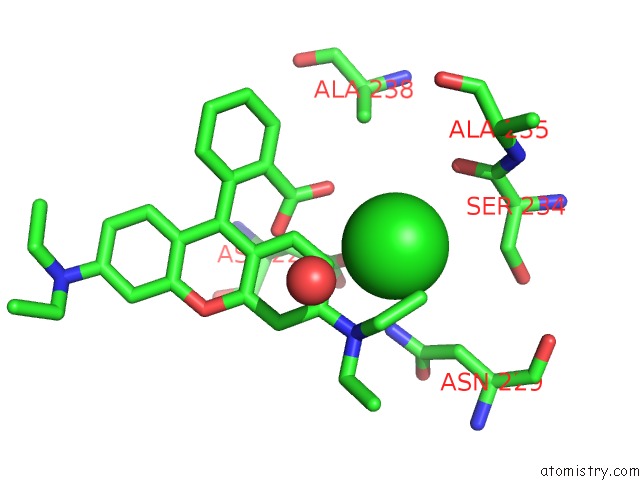
Mono view
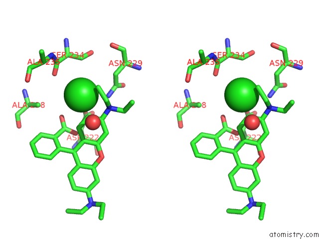
Stereo pair view
Mono view
Stereo pair view
A full contact list of Chlorine with other atoms in the Cl binding
site number 1 of Crystal Structure of Methylthioadenosine Phosphorylase in Complex with Rhodamine B within 5.0Å range:
|
Chlorine binding site 2 out of 2 in 4glj
Go back to
Chlorine binding site 2 out
of 2 in the Crystal Structure of Methylthioadenosine Phosphorylase in Complex with Rhodamine B
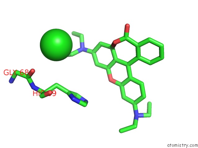
Mono view
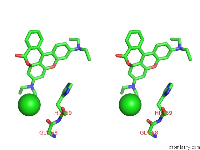
Stereo pair view
Mono view
Stereo pair view
A full contact list of Chlorine with other atoms in the Cl binding
site number 2 of Crystal Structure of Methylthioadenosine Phosphorylase in Complex with Rhodamine B within 5.0Å range:
|
Reference:
P.Bartasun,
H.Cieslinski,
A.Bujacz,
A.Wierzbicka-Wos,
J.Kur.
A Study on the Interaction of Rhodamine B with Methylthioadenosine Phosphorylase Protein Sourced From An Antarctic Soil Metagenomic Library. Plos One V. 8 55697 2013.
ISSN: ESSN 1932-6203
PubMed: 23383268
DOI: 10.1371/JOURNAL.PONE.0055697
Page generated: Sun Jul 21 14:45:59 2024
ISSN: ESSN 1932-6203
PubMed: 23383268
DOI: 10.1371/JOURNAL.PONE.0055697
Last articles
Cl in 2VJJCl in 2VJC
Cl in 2VJI
Cl in 2VJB
Cl in 2VJA
Cl in 2VIP
Cl in 2VJ3
Cl in 2VJ0
Cl in 2VIO
Cl in 2VIW