Chlorine »
PDB 4u3b-4ufx »
4ubs »
Chlorine in PDB 4ubs: The Crystal Structure of Cytochrome P450 105D7 From Streptomyces Avermitilis in Complex with Diclofenac
Enzymatic activity of The Crystal Structure of Cytochrome P450 105D7 From Streptomyces Avermitilis in Complex with Diclofenac
All present enzymatic activity of The Crystal Structure of Cytochrome P450 105D7 From Streptomyces Avermitilis in Complex with Diclofenac:
1.14.15.11;
1.14.15.11;
Protein crystallography data
The structure of The Crystal Structure of Cytochrome P450 105D7 From Streptomyces Avermitilis in Complex with Diclofenac, PDB code: 4ubs
was solved by
L.H.Xu,
H.Ikeda,
T.Arakawa,
T.Wakagi,
H.Shoun,
S.Fushinobu,
with X-Ray Crystallography technique. A brief refinement statistics is given in the table below:
| Resolution Low / High (Å) | 44.40 / 2.20 |
| Space group | P 31 2 1 |
| Cell size a, b, c (Å), α, β, γ (°) | 139.897, 139.897, 65.268, 90.00, 90.00, 120.00 |
| R / Rfree (%) | 17.7 / 22.8 |
Other elements in 4ubs:
The structure of The Crystal Structure of Cytochrome P450 105D7 From Streptomyces Avermitilis in Complex with Diclofenac also contains other interesting chemical elements:
| Iron | (Fe) | 1 atom |
Chlorine Binding Sites:
The binding sites of Chlorine atom in the The Crystal Structure of Cytochrome P450 105D7 From Streptomyces Avermitilis in Complex with Diclofenac
(pdb code 4ubs). This binding sites where shown within
5.0 Angstroms radius around Chlorine atom.
In total 4 binding sites of Chlorine where determined in the The Crystal Structure of Cytochrome P450 105D7 From Streptomyces Avermitilis in Complex with Diclofenac, PDB code: 4ubs:
Jump to Chlorine binding site number: 1; 2; 3; 4;
In total 4 binding sites of Chlorine where determined in the The Crystal Structure of Cytochrome P450 105D7 From Streptomyces Avermitilis in Complex with Diclofenac, PDB code: 4ubs:
Jump to Chlorine binding site number: 1; 2; 3; 4;
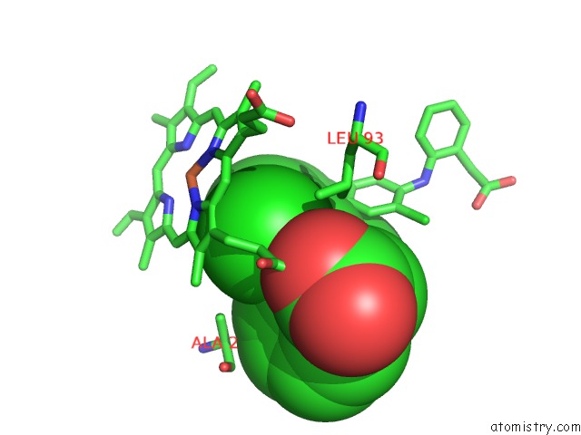
Chlorine binding site 1 out of 4 in 4ubs
Go back to
Chlorine binding site 1 out
of 4 in the The Crystal Structure of Cytochrome P450 105D7 From Streptomyces Avermitilis in Complex with Diclofenac
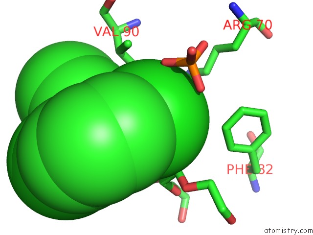
Mono view
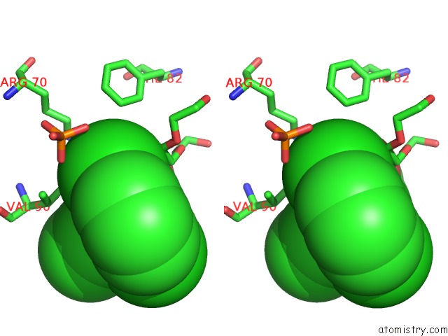
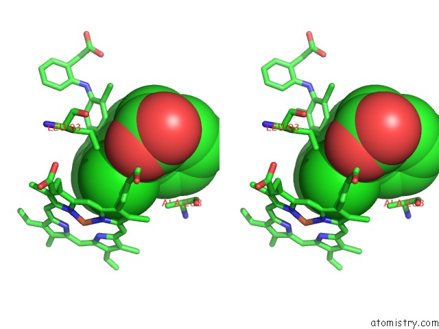
Stereo pair view
Mono view
Stereo pair view
A full contact list of Chlorine with other atoms in the Cl binding
site number 1 of The Crystal Structure of Cytochrome P450 105D7 From Streptomyces Avermitilis in Complex with Diclofenac within 5.0Å range:
|
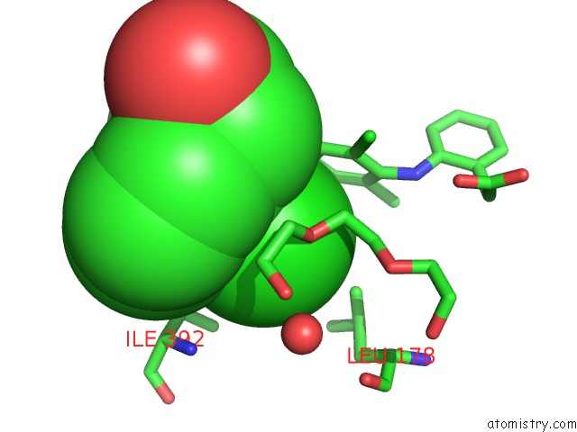
Chlorine binding site 2 out of 4 in 4ubs
Go back to
Chlorine binding site 2 out
of 4 in the The Crystal Structure of Cytochrome P450 105D7 From Streptomyces Avermitilis in Complex with Diclofenac
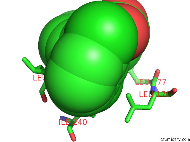
Mono view
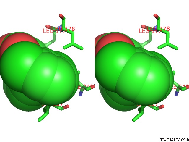
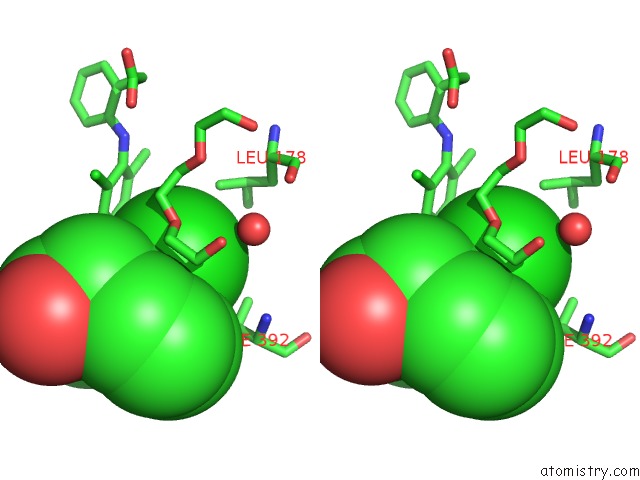
Stereo pair view
Mono view
Stereo pair view
A full contact list of Chlorine with other atoms in the Cl binding
site number 2 of The Crystal Structure of Cytochrome P450 105D7 From Streptomyces Avermitilis in Complex with Diclofenac within 5.0Å range:
|
Chlorine binding site 3 out of 4 in 4ubs
Go back to
Chlorine binding site 3 out
of 4 in the The Crystal Structure of Cytochrome P450 105D7 From Streptomyces Avermitilis in Complex with Diclofenac
Mono view
Stereo pair view
Mono view
Stereo pair view
A full contact list of Chlorine with other atoms in the Cl binding
site number 3 of The Crystal Structure of Cytochrome P450 105D7 From Streptomyces Avermitilis in Complex with Diclofenac within 5.0Å range:
|
Chlorine binding site 4 out of 4 in 4ubs
Go back to
Chlorine binding site 4 out
of 4 in the The Crystal Structure of Cytochrome P450 105D7 From Streptomyces Avermitilis in Complex with Diclofenac
Mono view
Stereo pair view
Mono view
Stereo pair view
A full contact list of Chlorine with other atoms in the Cl binding
site number 4 of The Crystal Structure of Cytochrome P450 105D7 From Streptomyces Avermitilis in Complex with Diclofenac within 5.0Å range:
|
Reference:
L.H.Xu,
H.Ikeda,
L.Liu,
T.Arakawa,
T.Wakagi,
H.Shoun,
S.Fushinobu.
Structural Basis For the 4'-Hydroxylation of Diclofenac By A Microbial Cytochrome P450 Monooxygenase. Appl.Microbiol.Biotechnol. 2014.
ISSN: ESSN 1432-0614
PubMed: 25341403
DOI: 10.1007/S00253-014-6148-Y
Page generated: Fri Jul 11 22:00:01 2025
ISSN: ESSN 1432-0614
PubMed: 25341403
DOI: 10.1007/S00253-014-6148-Y
Last articles
Cl in 5TOOCl in 5TO1
Cl in 5TNT
Cl in 5TM9
Cl in 5TML
Cl in 5TNO
Cl in 5TM7
Cl in 5TM4
Cl in 5TM3
Cl in 5TL4