Chlorine »
PDB 4z6v-4zg7 »
4z8n »
Chlorine in PDB 4z8n: Crystal Structure of the Erythrocyte-Binding Domain From Plasmodium Vivax Reticulocyte-Binding Protein 2A (PVRBP2A)
Protein crystallography data
The structure of Crystal Structure of the Erythrocyte-Binding Domain From Plasmodium Vivax Reticulocyte-Binding Protein 2A (PVRBP2A), PDB code: 4z8n
was solved by
J.Gruszczyk,
W.H.Tham,
with X-Ray Crystallography technique. A brief refinement statistics is given in the table below:
| Resolution Low / High (Å) | 43.84 / 2.12 |
| Space group | P 21 21 21 |
| Cell size a, b, c (Å), α, β, γ (°) | 58.790, 93.450, 126.720, 90.00, 90.00, 90.00 |
| R / Rfree (%) | 20.3 / 22.9 |
Other elements in 4z8n:
The structure of Crystal Structure of the Erythrocyte-Binding Domain From Plasmodium Vivax Reticulocyte-Binding Protein 2A (PVRBP2A) also contains other interesting chemical elements:
| Magnesium | (Mg) | 2 atoms |
| Calcium | (Ca) | 1 atom |
Chlorine Binding Sites:
The binding sites of Chlorine atom in the Crystal Structure of the Erythrocyte-Binding Domain From Plasmodium Vivax Reticulocyte-Binding Protein 2A (PVRBP2A)
(pdb code 4z8n). This binding sites where shown within
5.0 Angstroms radius around Chlorine atom.
In total only one binding site of Chlorine was determined in the Crystal Structure of the Erythrocyte-Binding Domain From Plasmodium Vivax Reticulocyte-Binding Protein 2A (PVRBP2A), PDB code: 4z8n:
In total only one binding site of Chlorine was determined in the Crystal Structure of the Erythrocyte-Binding Domain From Plasmodium Vivax Reticulocyte-Binding Protein 2A (PVRBP2A), PDB code: 4z8n:
Chlorine binding site 1 out of 1 in 4z8n
Go back to
Chlorine binding site 1 out
of 1 in the Crystal Structure of the Erythrocyte-Binding Domain From Plasmodium Vivax Reticulocyte-Binding Protein 2A (PVRBP2A)
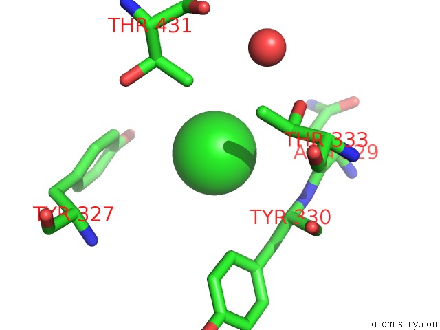
Mono view
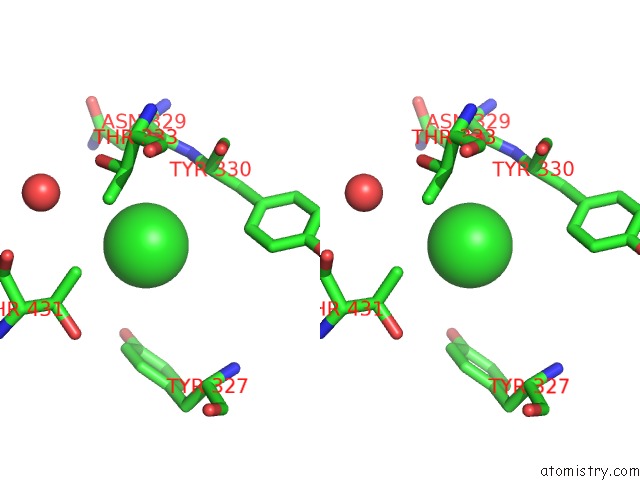
Stereo pair view
Mono view
Stereo pair view
A full contact list of Chlorine with other atoms in the Cl binding
site number 1 of Crystal Structure of the Erythrocyte-Binding Domain From Plasmodium Vivax Reticulocyte-Binding Protein 2A (PVRBP2A) within 5.0Å range:
|
Reference:
J.Gruszczyk,
N.T.Lim,
A.Arnott,
W.Q.He,
W.Nguitragool,
W.Roobsoong,
Y.F.Mok,
J.M.Murphy,
K.R.Smith,
S.Lee,
M.Bahlo,
I.Mueller,
A.E.Barry,
W.H.Tham.
Structurally Conserved Erythrocyte-Binding Domain in Plasmodium Provides A Versatile Scaffold For Alternate Receptor Engagement. Proc.Natl.Acad.Sci.Usa V. 113 E191 2016.
ISSN: ESSN 1091-6490
PubMed: 26715754
DOI: 10.1073/PNAS.1516512113
Page generated: Fri Jul 11 23:38:30 2025
ISSN: ESSN 1091-6490
PubMed: 26715754
DOI: 10.1073/PNAS.1516512113
Last articles
Cl in 7YNVCl in 7YNU
Cl in 7YCT
Cl in 7YP5
Cl in 7YOH
Cl in 7YK6
Cl in 7YIB
Cl in 7YD2
Cl in 7YI6
Cl in 7YD3