Chlorine »
PDB 5ofs-5on9 »
5omx »
Chlorine in PDB 5omx: X-Ray Structure of the H2A-N38C Nucleosome Core Particle
Protein crystallography data
The structure of X-Ray Structure of the H2A-N38C Nucleosome Core Particle, PDB code: 5omx
was solved by
T.D.Frouws,
T.J.Richmond,
with X-Ray Crystallography technique. A brief refinement statistics is given in the table below:
| Resolution Low / High (Å) | 47.14 / 2.32 |
| Space group | P 21 21 21 |
| Cell size a, b, c (Å), α, β, γ (°) | 106.753, 182.578, 109.645, 90.00, 90.00, 90.00 |
| R / Rfree (%) | 22.2 / 25.9 |
Other elements in 5omx:
The structure of X-Ray Structure of the H2A-N38C Nucleosome Core Particle also contains other interesting chemical elements:
| Manganese | (Mn) | 33 atoms |
Chlorine Binding Sites:
The binding sites of Chlorine atom in the X-Ray Structure of the H2A-N38C Nucleosome Core Particle
(pdb code 5omx). This binding sites where shown within
5.0 Angstroms radius around Chlorine atom.
In total 4 binding sites of Chlorine where determined in the X-Ray Structure of the H2A-N38C Nucleosome Core Particle, PDB code: 5omx:
Jump to Chlorine binding site number: 1; 2; 3; 4;
In total 4 binding sites of Chlorine where determined in the X-Ray Structure of the H2A-N38C Nucleosome Core Particle, PDB code: 5omx:
Jump to Chlorine binding site number: 1; 2; 3; 4;
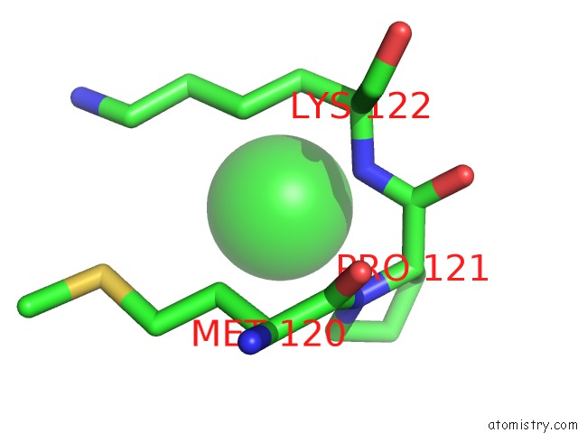
Chlorine binding site 1 out of 4 in 5omx
Go back to
Chlorine binding site 1 out
of 4 in the X-Ray Structure of the H2A-N38C Nucleosome Core Particle
Mono view

Stereo pair view
Mono view
Stereo pair view
A full contact list of Chlorine with other atoms in the Cl binding
site number 1 of X-Ray Structure of the H2A-N38C Nucleosome Core Particle within 5.0Å range:
|
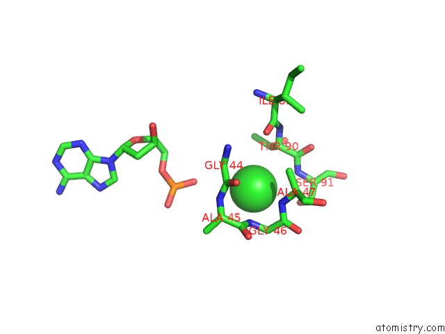
Chlorine binding site 2 out of 4 in 5omx
Go back to
Chlorine binding site 2 out
of 4 in the X-Ray Structure of the H2A-N38C Nucleosome Core Particle

Mono view

Stereo pair view
Mono view
Stereo pair view
A full contact list of Chlorine with other atoms in the Cl binding
site number 2 of X-Ray Structure of the H2A-N38C Nucleosome Core Particle within 5.0Å range:
|
Chlorine binding site 3 out of 4 in 5omx
Go back to
Chlorine binding site 3 out
of 4 in the X-Ray Structure of the H2A-N38C Nucleosome Core Particle

Mono view

Stereo pair view
Mono view
Stereo pair view
A full contact list of Chlorine with other atoms in the Cl binding
site number 3 of X-Ray Structure of the H2A-N38C Nucleosome Core Particle within 5.0Å range:
|
Chlorine binding site 4 out of 4 in 5omx
Go back to
Chlorine binding site 4 out
of 4 in the X-Ray Structure of the H2A-N38C Nucleosome Core Particle
Mono view

Stereo pair view
Mono view
Stereo pair view
A full contact list of Chlorine with other atoms in the Cl binding
site number 4 of X-Ray Structure of the H2A-N38C Nucleosome Core Particle within 5.0Å range:
|
Reference:
T.D.Frouws,
P.D.Barth,
T.J.Richmond.
Site-Specific Disulfide Crosslinked Nucleosomes with Enhanced Stability. J. Mol. Biol. V. 430 45 2018.
ISSN: ESSN 1089-8638
PubMed: 29113904
DOI: 10.1016/J.JMB.2017.10.029
Page generated: Sat Jul 12 06:49:48 2025
ISSN: ESSN 1089-8638
PubMed: 29113904
DOI: 10.1016/J.JMB.2017.10.029
Last articles
Cl in 8AHQCl in 8AHY
Cl in 8AHO
Cl in 8AFN
Cl in 8AGA
Cl in 8AFJ
Cl in 8AF1
Cl in 8AEU
Cl in 8AEP
Cl in 8AEM