Chlorine »
PDB 5vc5-5vm6 »
5vd5 »
Chlorine in PDB 5vd5: Crystal Structure of Human WEE1 Kinase Domain in Complex with Rac-IV- 050, A MK1775 Analougue
Enzymatic activity of Crystal Structure of Human WEE1 Kinase Domain in Complex with Rac-IV- 050, A MK1775 Analougue
All present enzymatic activity of Crystal Structure of Human WEE1 Kinase Domain in Complex with Rac-IV- 050, A MK1775 Analougue:
2.7.10.2;
2.7.10.2;
Protein crystallography data
The structure of Crystal Structure of Human WEE1 Kinase Domain in Complex with Rac-IV- 050, A MK1775 Analougue, PDB code: 5vd5
was solved by
J.-Y.Zhu,
E.Schonbrunn,
with X-Ray Crystallography technique. A brief refinement statistics is given in the table below:
| Resolution Low / High (Å) | 36.43 / 2.05 |
| Space group | P 1 21 1 |
| Cell size a, b, c (Å), α, β, γ (°) | 50.240, 44.580, 64.650, 90.00, 102.17, 90.00 |
| R / Rfree (%) | 19.5 / 23.4 |
Chlorine Binding Sites:
The binding sites of Chlorine atom in the Crystal Structure of Human WEE1 Kinase Domain in Complex with Rac-IV- 050, A MK1775 Analougue
(pdb code 5vd5). This binding sites where shown within
5.0 Angstroms radius around Chlorine atom.
In total only one binding site of Chlorine was determined in the Crystal Structure of Human WEE1 Kinase Domain in Complex with Rac-IV- 050, A MK1775 Analougue, PDB code: 5vd5:
In total only one binding site of Chlorine was determined in the Crystal Structure of Human WEE1 Kinase Domain in Complex with Rac-IV- 050, A MK1775 Analougue, PDB code: 5vd5:
Chlorine binding site 1 out of 1 in 5vd5
Go back to
Chlorine binding site 1 out
of 1 in the Crystal Structure of Human WEE1 Kinase Domain in Complex with Rac-IV- 050, A MK1775 Analougue
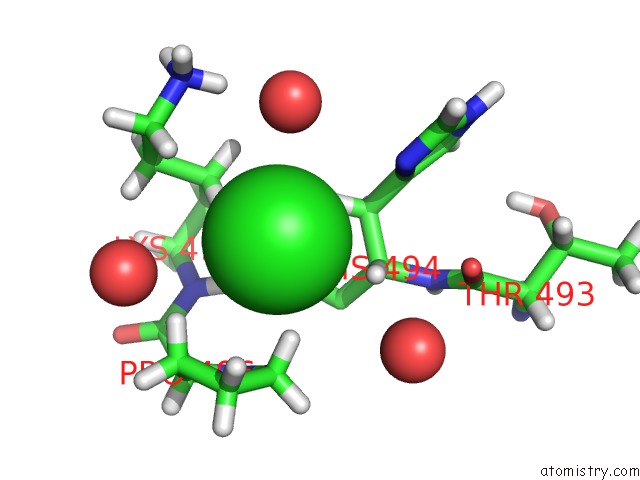
Mono view
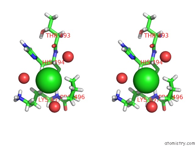
Stereo pair view
Mono view
Stereo pair view
A full contact list of Chlorine with other atoms in the Cl binding
site number 1 of Crystal Structure of Human WEE1 Kinase Domain in Complex with Rac-IV- 050, A MK1775 Analougue within 5.0Å range:
|
Reference:
J.-Y.Zhu,
E.Schonbrunn.
Structural Basis of Wee Family Kinase Inhibition By Small Molecules To Be Published.
Page generated: Sat Jul 12 09:51:21 2025
Last articles
Cl in 6HT2Cl in 6HTM
Cl in 6HTY
Cl in 6HTP
Cl in 6HTI
Cl in 6HTG
Cl in 6HTD
Cl in 6HTC
Cl in 6HTB
Cl in 6HSM