Chlorine »
PDB 5wkx-5ws6 »
5wnl »
Chlorine in PDB 5wnl: Crystal Structure of Murine Receptor-Interacting Protein 4 (RIPK4) D143N Bound to Staurosporine
Enzymatic activity of Crystal Structure of Murine Receptor-Interacting Protein 4 (RIPK4) D143N Bound to Staurosporine
All present enzymatic activity of Crystal Structure of Murine Receptor-Interacting Protein 4 (RIPK4) D143N Bound to Staurosporine:
2.7.11.1;
2.7.11.1;
Protein crystallography data
The structure of Crystal Structure of Murine Receptor-Interacting Protein 4 (RIPK4) D143N Bound to Staurosporine, PDB code: 5wnl
was solved by
C.S.Huang,
S.G.Hymowitz,
with X-Ray Crystallography technique. A brief refinement statistics is given in the table below:
| Resolution Low / High (Å) | 30.00 / 2.50 |
| Space group | I 21 21 21 |
| Cell size a, b, c (Å), α, β, γ (°) | 70.158, 109.563, 146.221, 90.00, 90.00, 90.00 |
| R / Rfree (%) | 22.3 / 25.6 |
Chlorine Binding Sites:
The binding sites of Chlorine atom in the Crystal Structure of Murine Receptor-Interacting Protein 4 (RIPK4) D143N Bound to Staurosporine
(pdb code 5wnl). This binding sites where shown within
5.0 Angstroms radius around Chlorine atom.
In total only one binding site of Chlorine was determined in the Crystal Structure of Murine Receptor-Interacting Protein 4 (RIPK4) D143N Bound to Staurosporine, PDB code: 5wnl:
In total only one binding site of Chlorine was determined in the Crystal Structure of Murine Receptor-Interacting Protein 4 (RIPK4) D143N Bound to Staurosporine, PDB code: 5wnl:
Chlorine binding site 1 out of 1 in 5wnl
Go back to
Chlorine binding site 1 out
of 1 in the Crystal Structure of Murine Receptor-Interacting Protein 4 (RIPK4) D143N Bound to Staurosporine
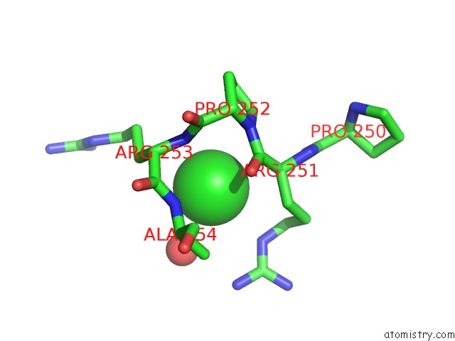
Mono view
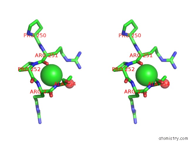
Stereo pair view
Mono view
Stereo pair view
A full contact list of Chlorine with other atoms in the Cl binding
site number 1 of Crystal Structure of Murine Receptor-Interacting Protein 4 (RIPK4) D143N Bound to Staurosporine within 5.0Å range:
|
Reference:
C.S.Huang,
N.Oberbeck,
Y.C.Hsiao,
P.Liu,
A.R.Johnson,
V.M.Dixit,
S.G.Hymowitz.
Crystal Structure of RIPK4 Reveals Dimerization-Dependent Kinase Activity. Structure V. 26 767 2018.
ISSN: ISSN 1878-4186
PubMed: 29706531
DOI: 10.1016/J.STR.2018.04.002
Page generated: Sat Jul 12 10:22:33 2025
ISSN: ISSN 1878-4186
PubMed: 29706531
DOI: 10.1016/J.STR.2018.04.002
Last articles
Cl in 6H5XCl in 6H57
Cl in 6H5W
Cl in 6H5V
Cl in 6H5T
Cl in 6H54
Cl in 6H4Y
Cl in 6H4M
Cl in 6H4Z
Cl in 6H4X