Chlorine »
PDB 7mde-7mnd »
7mi0 »
Chlorine in PDB 7mi0: Crystal Structure of Glycosyltransferase From Rickettsia Africae Esf-5
Protein crystallography data
The structure of Crystal Structure of Glycosyltransferase From Rickettsia Africae Esf-5, PDB code: 7mi0
was solved by
Seattle Structural Genomics Center For Infectious Disease (Ssgcid),
with X-Ray Crystallography technique. A brief refinement statistics is given in the table below:
| Resolution Low / High (Å) | 47.49 / 2.90 |
| Space group | P 65 2 2 |
| Cell size a, b, c (Å), α, β, γ (°) | 111.27, 111.27, 163.74, 90, 90, 120 |
| R / Rfree (%) | 19.4 / 23.6 |
Chlorine Binding Sites:
The binding sites of Chlorine atom in the Crystal Structure of Glycosyltransferase From Rickettsia Africae Esf-5
(pdb code 7mi0). This binding sites where shown within
5.0 Angstroms radius around Chlorine atom.
In total 4 binding sites of Chlorine where determined in the Crystal Structure of Glycosyltransferase From Rickettsia Africae Esf-5, PDB code: 7mi0:
Jump to Chlorine binding site number: 1; 2; 3; 4;
In total 4 binding sites of Chlorine where determined in the Crystal Structure of Glycosyltransferase From Rickettsia Africae Esf-5, PDB code: 7mi0:
Jump to Chlorine binding site number: 1; 2; 3; 4;
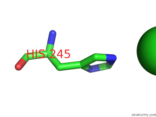
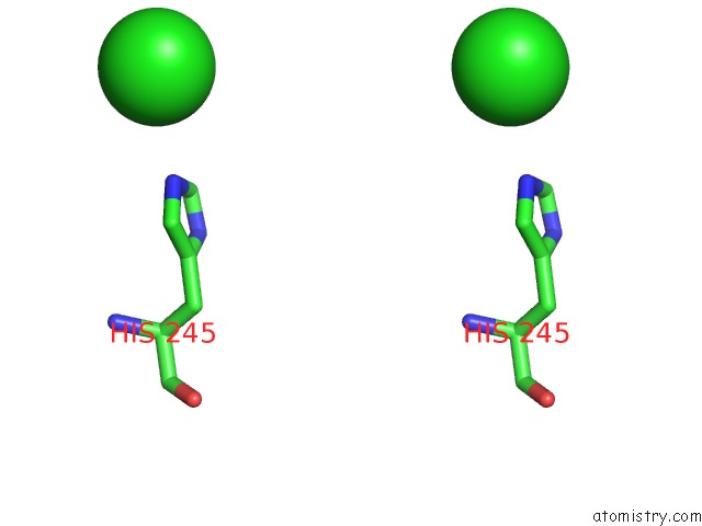
Chlorine binding site 1 out of 4 in 7mi0
Go back to
Chlorine binding site 1 out
of 4 in the Crystal Structure of Glycosyltransferase From Rickettsia Africae Esf-5

Mono view

Stereo pair view
Mono view
Stereo pair view
A full contact list of Chlorine with other atoms in the Cl binding
site number 1 of Crystal Structure of Glycosyltransferase From Rickettsia Africae Esf-5 within 5.0Å range:
|
Chlorine binding site 2 out of 4 in 7mi0
Go back to
Chlorine binding site 2 out
of 4 in the Crystal Structure of Glycosyltransferase From Rickettsia Africae Esf-5

Mono view

Stereo pair view
Mono view
Stereo pair view
A full contact list of Chlorine with other atoms in the Cl binding
site number 2 of Crystal Structure of Glycosyltransferase From Rickettsia Africae Esf-5 within 5.0Å range:
|
Chlorine binding site 3 out of 4 in 7mi0
Go back to
Chlorine binding site 3 out
of 4 in the Crystal Structure of Glycosyltransferase From Rickettsia Africae Esf-5
Mono view
Stereo pair view
Mono view
Stereo pair view
A full contact list of Chlorine with other atoms in the Cl binding
site number 3 of Crystal Structure of Glycosyltransferase From Rickettsia Africae Esf-5 within 5.0Å range:
|
Chlorine binding site 4 out of 4 in 7mi0
Go back to
Chlorine binding site 4 out
of 4 in the Crystal Structure of Glycosyltransferase From Rickettsia Africae Esf-5

Mono view

Stereo pair view
Mono view
Stereo pair view
A full contact list of Chlorine with other atoms in the Cl binding
site number 4 of Crystal Structure of Glycosyltransferase From Rickettsia Africae Esf-5 within 5.0Å range:
|
Reference:
J.Abendroth,
D.R.Davies,
D.D.Lorimer,
P.S.Horanyi,
T.E.Edwards.
Crystal Structure of Glycosyltransferase From Rickettsia Africae Esf-5 To Be Published.
Page generated: Sun Jul 13 04:07:08 2025
Last articles
Cl in 8AHZCl in 8AHQ
Cl in 8AHY
Cl in 8AHO
Cl in 8AFN
Cl in 8AGA
Cl in 8AFJ
Cl in 8AF1
Cl in 8AEU
Cl in 8AEP