Chlorine »
PDB 3lnz-3lz0 »
3ls2 »
Chlorine in PDB 3ls2: Crystal Structure of An S-Formylglutathione Hydrolase From Pseudoalteromonas Haloplanktis TAC125
Protein crystallography data
The structure of Crystal Structure of An S-Formylglutathione Hydrolase From Pseudoalteromonas Haloplanktis TAC125, PDB code: 3ls2
was solved by
V.Alterio,
G.De Simone,
with X-Ray Crystallography technique. A brief refinement statistics is given in the table below:
| Resolution Low / High (Å) | 20.00 / 2.20 |
| Space group | P 21 21 21 |
| Cell size a, b, c (Å), α, β, γ (°) | 49.490, 129.750, 152.670, 90.00, 90.00, 90.00 |
| R / Rfree (%) | 16.1 / 20.5 |
Chlorine Binding Sites:
The binding sites of Chlorine atom in the Crystal Structure of An S-Formylglutathione Hydrolase From Pseudoalteromonas Haloplanktis TAC125
(pdb code 3ls2). This binding sites where shown within
5.0 Angstroms radius around Chlorine atom.
In total 4 binding sites of Chlorine where determined in the Crystal Structure of An S-Formylglutathione Hydrolase From Pseudoalteromonas Haloplanktis TAC125, PDB code: 3ls2:
Jump to Chlorine binding site number: 1; 2; 3; 4;
In total 4 binding sites of Chlorine where determined in the Crystal Structure of An S-Formylglutathione Hydrolase From Pseudoalteromonas Haloplanktis TAC125, PDB code: 3ls2:
Jump to Chlorine binding site number: 1; 2; 3; 4;
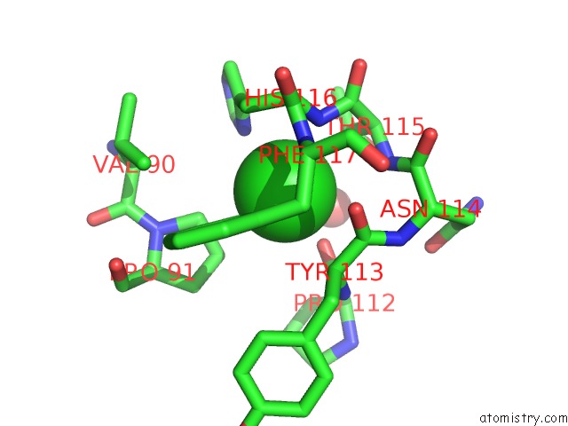
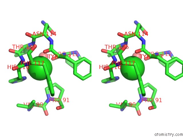
Chlorine binding site 1 out of 4 in 3ls2
Go back to
Chlorine binding site 1 out
of 4 in the Crystal Structure of An S-Formylglutathione Hydrolase From Pseudoalteromonas Haloplanktis TAC125
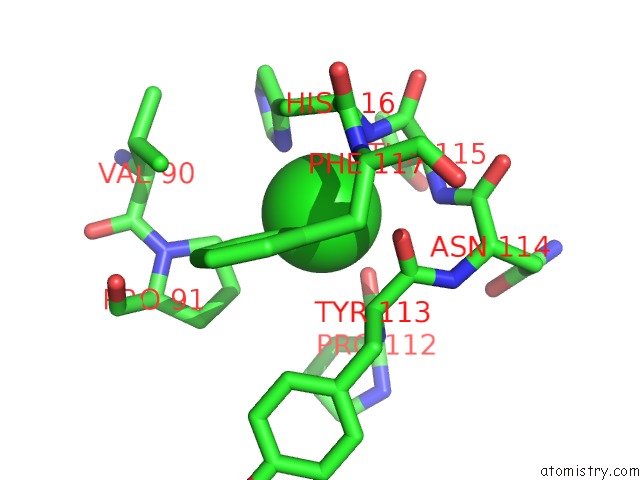
Mono view
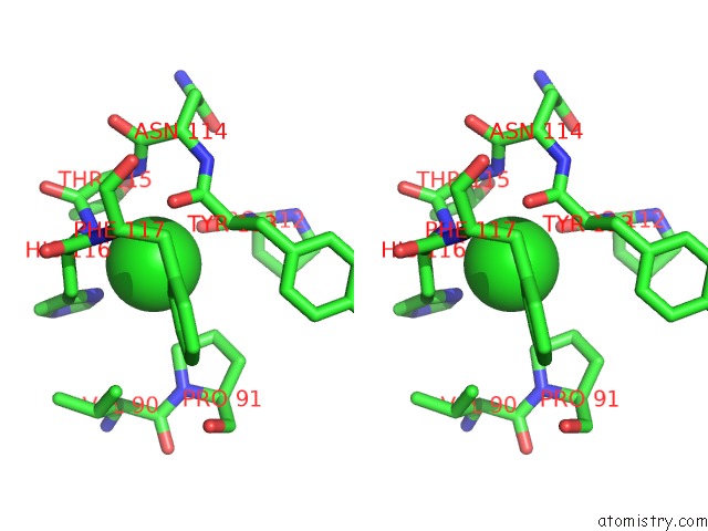
Stereo pair view
Mono view
Stereo pair view
A full contact list of Chlorine with other atoms in the Cl binding
site number 1 of Crystal Structure of An S-Formylglutathione Hydrolase From Pseudoalteromonas Haloplanktis TAC125 within 5.0Å range:
|
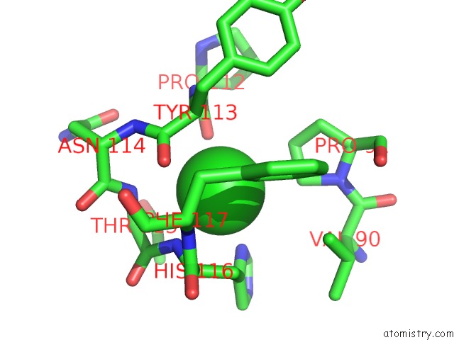
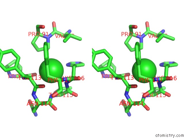
Chlorine binding site 2 out of 4 in 3ls2
Go back to
Chlorine binding site 2 out
of 4 in the Crystal Structure of An S-Formylglutathione Hydrolase From Pseudoalteromonas Haloplanktis TAC125
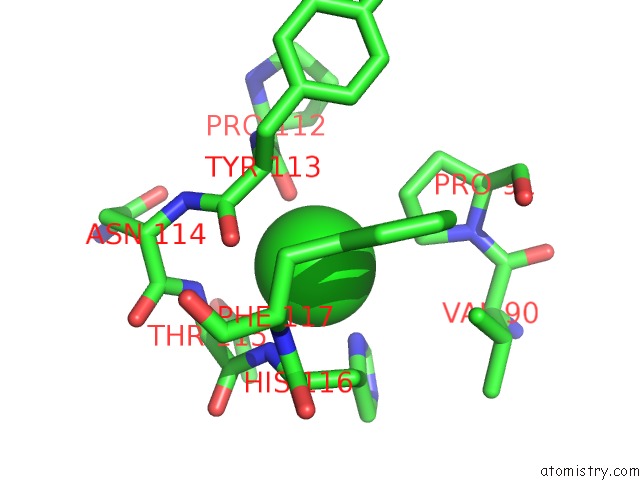
Mono view
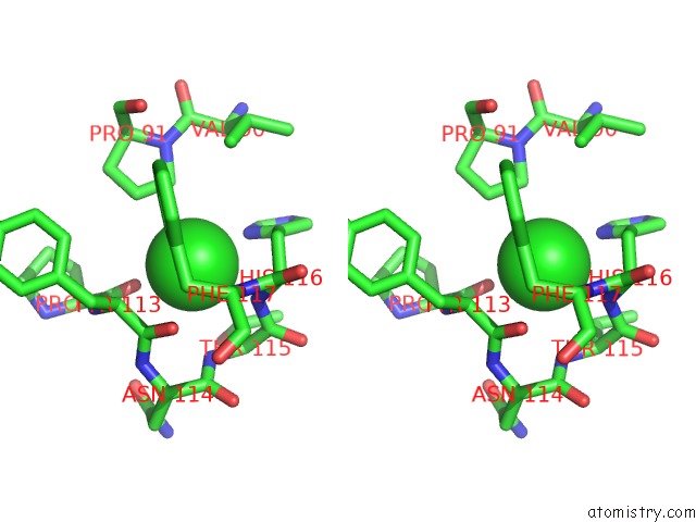
Stereo pair view
Mono view
Stereo pair view
A full contact list of Chlorine with other atoms in the Cl binding
site number 2 of Crystal Structure of An S-Formylglutathione Hydrolase From Pseudoalteromonas Haloplanktis TAC125 within 5.0Å range:
|
Chlorine binding site 3 out of 4 in 3ls2
Go back to
Chlorine binding site 3 out
of 4 in the Crystal Structure of An S-Formylglutathione Hydrolase From Pseudoalteromonas Haloplanktis TAC125
Mono view
Stereo pair view
Mono view
Stereo pair view
A full contact list of Chlorine with other atoms in the Cl binding
site number 3 of Crystal Structure of An S-Formylglutathione Hydrolase From Pseudoalteromonas Haloplanktis TAC125 within 5.0Å range:
|
Chlorine binding site 4 out of 4 in 3ls2
Go back to
Chlorine binding site 4 out
of 4 in the Crystal Structure of An S-Formylglutathione Hydrolase From Pseudoalteromonas Haloplanktis TAC125
Mono view
Stereo pair view
Mono view
Stereo pair view
A full contact list of Chlorine with other atoms in the Cl binding
site number 4 of Crystal Structure of An S-Formylglutathione Hydrolase From Pseudoalteromonas Haloplanktis TAC125 within 5.0Å range:
|
Reference:
V.Alterio,
V.Aurilia,
A.Romanelli,
A.Parracino,
M.Saviano,
S.D'auria,
G.De Simone.
Crystal Structure of An S-Formylglutathione Hydrolase From Pseudoalteromonas Haloplanktis TAC125. Biopolymers V. 93 669 2010.
ISSN: ISSN 0006-3525
PubMed: 20209484
DOI: 10.1002/BIP.21420
Page generated: Fri Jul 11 07:33:26 2025
ISSN: ISSN 0006-3525
PubMed: 20209484
DOI: 10.1002/BIP.21420
Last articles
Cl in 3NB5Cl in 3NA6
Cl in 3N9J
Cl in 3N9E
Cl in 3N9C
Cl in 3N9B
Cl in 3N8Y
Cl in 3N9A
Cl in 3N8U
Cl in 3N7T