Chlorine »
PDB 4aj2-4aqd »
4akd »
Chlorine in PDB 4akd: High Resolution Structure of Mannose Binding Lectin From Champedak (Cmb)
Protein crystallography data
The structure of High Resolution Structure of Mannose Binding Lectin From Champedak (Cmb), PDB code: 4akd
was solved by
M.Gabrielsen,
P.S.Abdul-Rahman,
S.Othman,
O.H.Hashim,
R.J.Cogdell,
with X-Ray Crystallography technique. A brief refinement statistics is given in the table below:
| Resolution Low / High (Å) | 47.69 / 2.10 |
| Space group | P 21 21 21 |
| Cell size a, b, c (Å), α, β, γ (°) | 76.887, 86.222, 95.373, 90.00, 90.00, 90.00 |
| R / Rfree (%) | 19.34 / 23.57 |
Other elements in 4akd:
The structure of High Resolution Structure of Mannose Binding Lectin From Champedak (Cmb) also contains other interesting chemical elements:
| Cadmium | (Cd) | 8 atoms |
Chlorine Binding Sites:
The binding sites of Chlorine atom in the High Resolution Structure of Mannose Binding Lectin From Champedak (Cmb)
(pdb code 4akd). This binding sites where shown within
5.0 Angstroms radius around Chlorine atom.
In total 7 binding sites of Chlorine where determined in the High Resolution Structure of Mannose Binding Lectin From Champedak (Cmb), PDB code: 4akd:
Jump to Chlorine binding site number: 1; 2; 3; 4; 5; 6; 7;
In total 7 binding sites of Chlorine where determined in the High Resolution Structure of Mannose Binding Lectin From Champedak (Cmb), PDB code: 4akd:
Jump to Chlorine binding site number: 1; 2; 3; 4; 5; 6; 7;
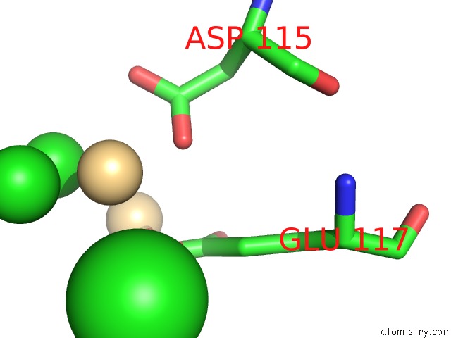
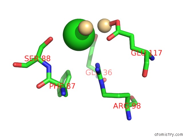
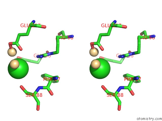
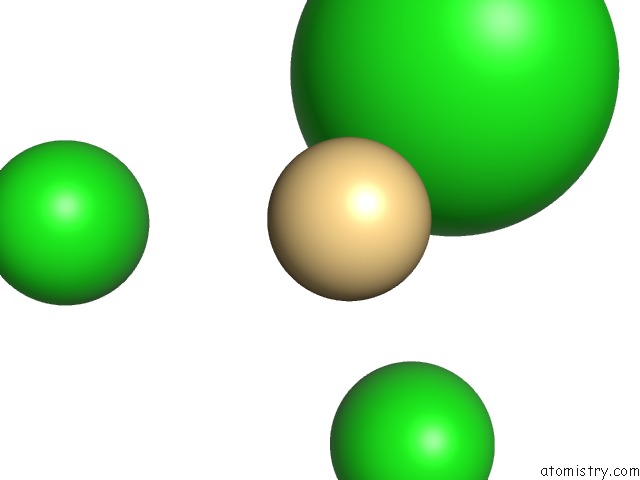
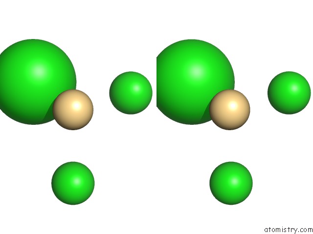
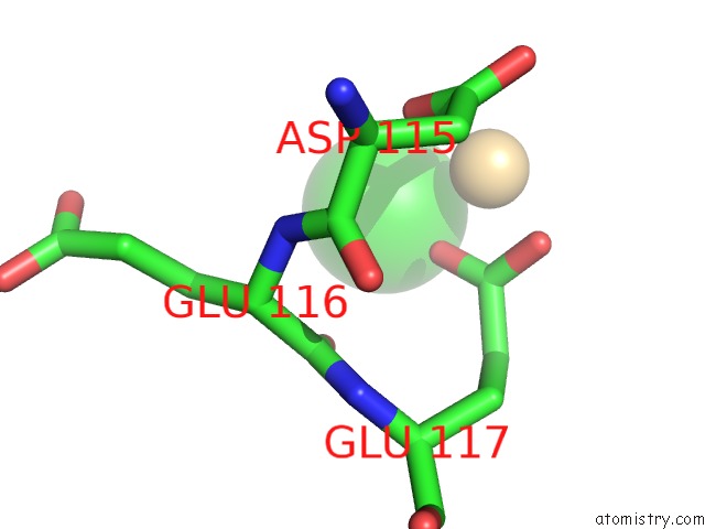
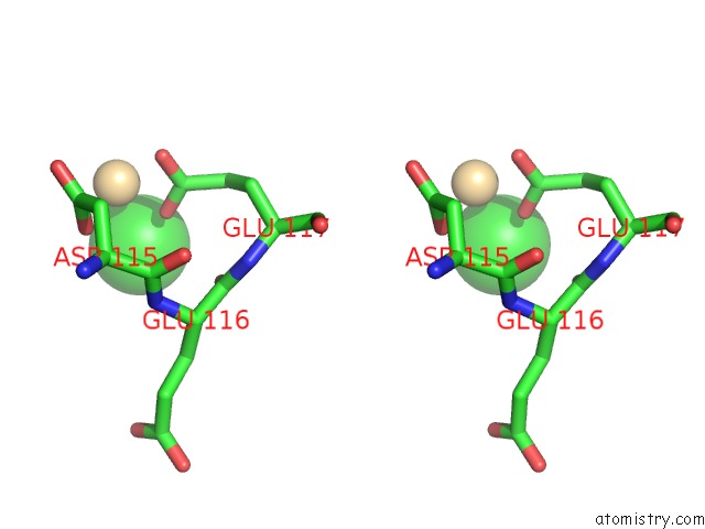
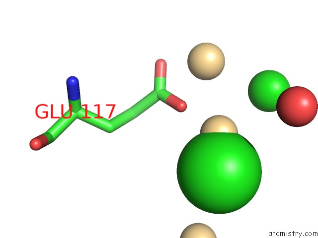
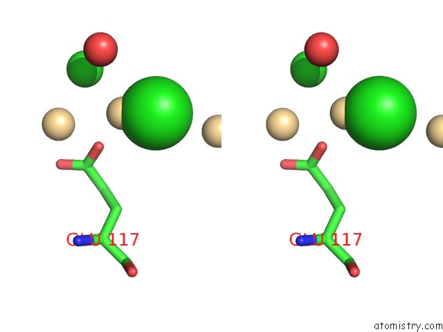
Chlorine binding site 1 out of 7 in 4akd
Go back to
Chlorine binding site 1 out
of 7 in the High Resolution Structure of Mannose Binding Lectin From Champedak (Cmb)
Mono view
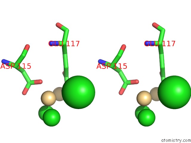
Stereo pair view
Mono view
Stereo pair view
A full contact list of Chlorine with other atoms in the Cl binding
site number 1 of High Resolution Structure of Mannose Binding Lectin From Champedak (Cmb) within 5.0Å range:
|
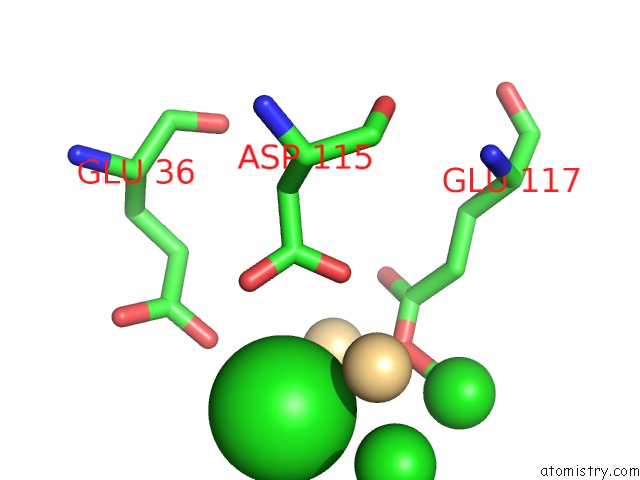
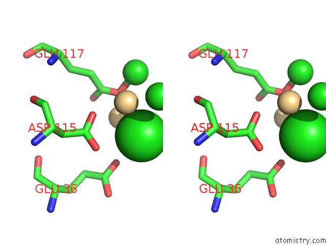
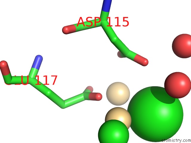
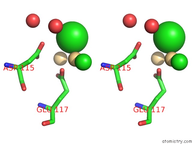
Chlorine binding site 2 out of 7 in 4akd
Go back to
Chlorine binding site 2 out
of 7 in the High Resolution Structure of Mannose Binding Lectin From Champedak (Cmb)
Mono view
Stereo pair view
Mono view
Stereo pair view
A full contact list of Chlorine with other atoms in the Cl binding
site number 2 of High Resolution Structure of Mannose Binding Lectin From Champedak (Cmb) within 5.0Å range:
|
Chlorine binding site 3 out of 7 in 4akd
Go back to
Chlorine binding site 3 out
of 7 in the High Resolution Structure of Mannose Binding Lectin From Champedak (Cmb)
Mono view
Stereo pair view
Mono view
Stereo pair view
A full contact list of Chlorine with other atoms in the Cl binding
site number 3 of High Resolution Structure of Mannose Binding Lectin From Champedak (Cmb) within 5.0Å range:
|
Chlorine binding site 4 out of 7 in 4akd
Go back to
Chlorine binding site 4 out
of 7 in the High Resolution Structure of Mannose Binding Lectin From Champedak (Cmb)
Mono view
Stereo pair view
Mono view
Stereo pair view
A full contact list of Chlorine with other atoms in the Cl binding
site number 4 of High Resolution Structure of Mannose Binding Lectin From Champedak (Cmb) within 5.0Å range:
|
Chlorine binding site 5 out of 7 in 4akd
Go back to
Chlorine binding site 5 out
of 7 in the High Resolution Structure of Mannose Binding Lectin From Champedak (Cmb)
Mono view
Stereo pair view
Mono view
Stereo pair view
A full contact list of Chlorine with other atoms in the Cl binding
site number 5 of High Resolution Structure of Mannose Binding Lectin From Champedak (Cmb) within 5.0Å range:
|
Chlorine binding site 6 out of 7 in 4akd
Go back to
Chlorine binding site 6 out
of 7 in the High Resolution Structure of Mannose Binding Lectin From Champedak (Cmb)
Mono view
Stereo pair view
Mono view
Stereo pair view
A full contact list of Chlorine with other atoms in the Cl binding
site number 6 of High Resolution Structure of Mannose Binding Lectin From Champedak (Cmb) within 5.0Å range:
|
Chlorine binding site 7 out of 7 in 4akd
Go back to
Chlorine binding site 7 out
of 7 in the High Resolution Structure of Mannose Binding Lectin From Champedak (Cmb)
Mono view
Stereo pair view
Mono view
Stereo pair view
A full contact list of Chlorine with other atoms in the Cl binding
site number 7 of High Resolution Structure of Mannose Binding Lectin From Champedak (Cmb) within 5.0Å range:
|
Reference:
M.Gabrielsen,
P.S.Abdul-Rahman,
S.Othman,
O.H.Hashim,
R.J.Cogdell.
Structures and Binding Specificity of Galactose- and Mannose-Binding Lectins From Champedak: Differences From Jackfruit Lectins Acta Crystallogr.,Sect.F V. 70 709 2014.
ISSN: ISSN 1744-3091
PubMed: 24915077
DOI: 10.1107/S2053230X14008966
Page generated: Fri Jul 11 12:52:25 2025
ISSN: ISSN 1744-3091
PubMed: 24915077
DOI: 10.1107/S2053230X14008966
Last articles
Mo in 4WN9Mo in 4WES
Mo in 4USA
Mo in 4US9
Mo in 4UHX
Mo in 4US8
Mo in 4NDP
Mo in 4UHW
Mo in 4TKV
Mo in 4TKU