Chlorine »
PDB 4aj2-4aqd »
4ap6 »
Chlorine in PDB 4ap6: Crystal Structure of Human POFUT2 E54A Mutant in Complex with Gdp- Fucose
Enzymatic activity of Crystal Structure of Human POFUT2 E54A Mutant in Complex with Gdp- Fucose
All present enzymatic activity of Crystal Structure of Human POFUT2 E54A Mutant in Complex with Gdp- Fucose:
2.4.1.221;
2.4.1.221;
Protein crystallography data
The structure of Crystal Structure of Human POFUT2 E54A Mutant in Complex with Gdp- Fucose, PDB code: 4ap6
was solved by
C.Chen,
J.J.Keusch,
D.Klein,
D.Hess,
J.Hofsteenge,
H.Gut,
with X-Ray Crystallography technique. A brief refinement statistics is given in the table below:
| Resolution Low / High (Å) | 39.686 / 3.401 |
| Space group | P 32 2 1 |
| Cell size a, b, c (Å), α, β, γ (°) | 153.010, 153.010, 185.680, 90.00, 90.00, 120.00 |
| R / Rfree (%) | 19.24 / 23.8 |
Chlorine Binding Sites:
The binding sites of Chlorine atom in the Crystal Structure of Human POFUT2 E54A Mutant in Complex with Gdp- Fucose
(pdb code 4ap6). This binding sites where shown within
5.0 Angstroms radius around Chlorine atom.
In total 4 binding sites of Chlorine where determined in the Crystal Structure of Human POFUT2 E54A Mutant in Complex with Gdp- Fucose, PDB code: 4ap6:
Jump to Chlorine binding site number: 1; 2; 3; 4;
In total 4 binding sites of Chlorine where determined in the Crystal Structure of Human POFUT2 E54A Mutant in Complex with Gdp- Fucose, PDB code: 4ap6:
Jump to Chlorine binding site number: 1; 2; 3; 4;
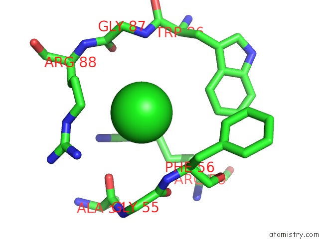
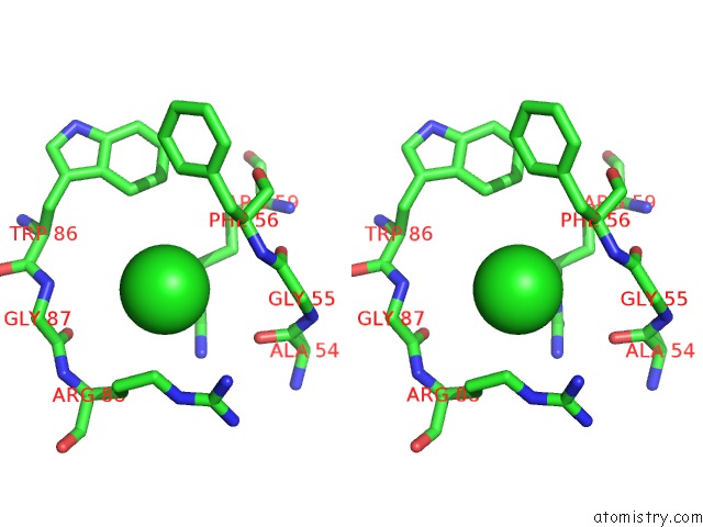
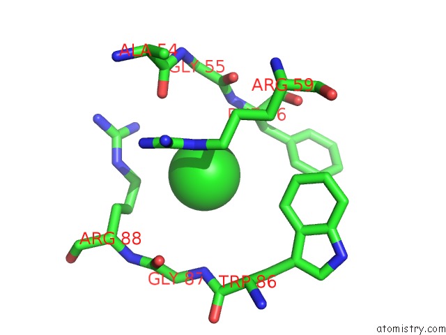
Chlorine binding site 1 out of 4 in 4ap6
Go back to
Chlorine binding site 1 out
of 4 in the Crystal Structure of Human POFUT2 E54A Mutant in Complex with Gdp- Fucose
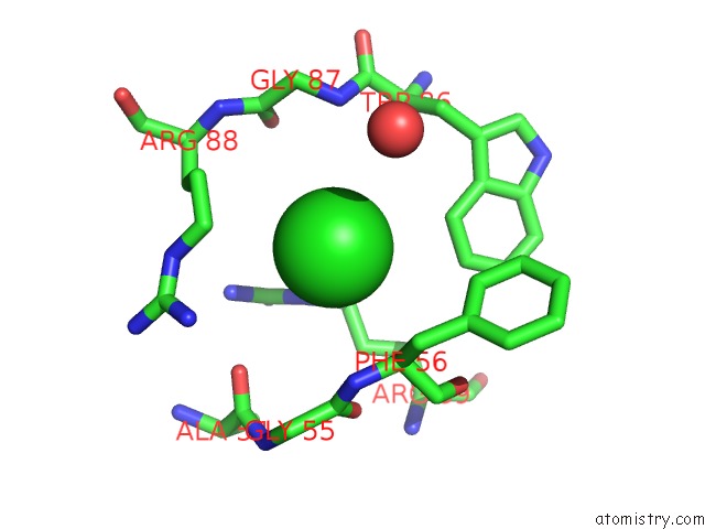
Mono view
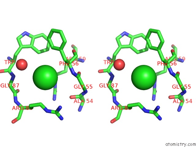
Stereo pair view
Mono view
Stereo pair view
A full contact list of Chlorine with other atoms in the Cl binding
site number 1 of Crystal Structure of Human POFUT2 E54A Mutant in Complex with Gdp- Fucose within 5.0Å range:
|
Chlorine binding site 2 out of 4 in 4ap6
Go back to
Chlorine binding site 2 out
of 4 in the Crystal Structure of Human POFUT2 E54A Mutant in Complex with Gdp- Fucose
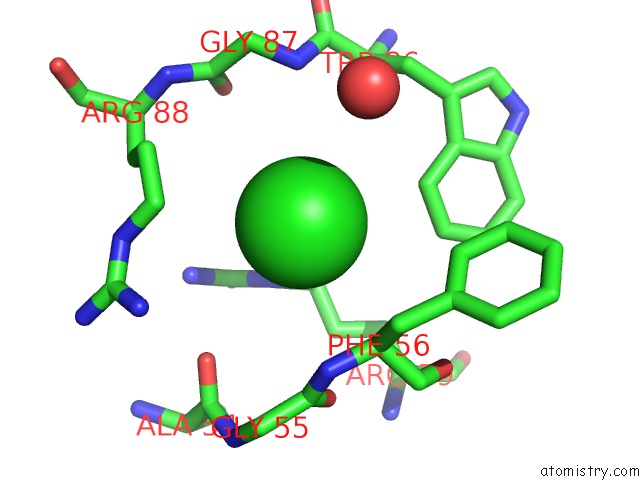
Mono view
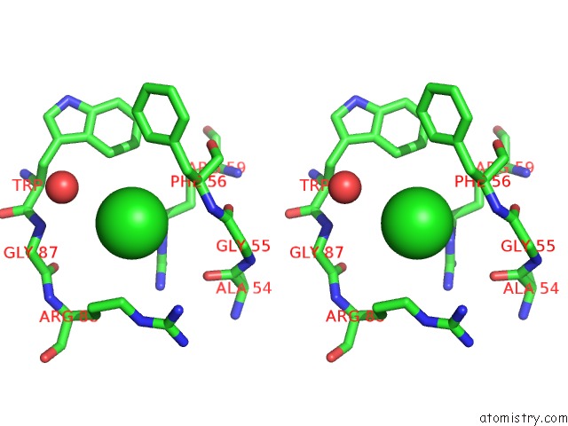
Stereo pair view
Mono view
Stereo pair view
A full contact list of Chlorine with other atoms in the Cl binding
site number 2 of Crystal Structure of Human POFUT2 E54A Mutant in Complex with Gdp- Fucose within 5.0Å range:
|
Chlorine binding site 3 out of 4 in 4ap6
Go back to
Chlorine binding site 3 out
of 4 in the Crystal Structure of Human POFUT2 E54A Mutant in Complex with Gdp- Fucose
Mono view
Stereo pair view
Mono view
Stereo pair view
A full contact list of Chlorine with other atoms in the Cl binding
site number 3 of Crystal Structure of Human POFUT2 E54A Mutant in Complex with Gdp- Fucose within 5.0Å range:
|
Chlorine binding site 4 out of 4 in 4ap6
Go back to
Chlorine binding site 4 out
of 4 in the Crystal Structure of Human POFUT2 E54A Mutant in Complex with Gdp- Fucose
Mono view

Stereo pair view
Mono view
Stereo pair view
A full contact list of Chlorine with other atoms in the Cl binding
site number 4 of Crystal Structure of Human POFUT2 E54A Mutant in Complex with Gdp- Fucose within 5.0Å range:
|
Reference:
C.I.Chen,
J.J.Keusch,
D.Klein,
D.Hess,
J.Hofsteenge,
H.Gut.
Structure of Human POFUT2: Insights Into Thrombospondin Type 1 Repeat Fold and O-Fucosylation. Embo J. V. 31 3183 2012.
ISSN: ISSN 0261-4189
PubMed: 22588082
DOI: 10.1038/EMBOJ.2012.143
Page generated: Fri Jul 11 12:57:45 2025
ISSN: ISSN 0261-4189
PubMed: 22588082
DOI: 10.1038/EMBOJ.2012.143
Last articles
K in 5BJPK in 5B2S
K in 5B2R
K in 5B2T
K in 5BJO
K in 5AW9
K in 5AYN
K in 5AYO
K in 5AXG
K in 5AW6