Chlorine »
PDB 4aj2-4aqd »
4apu »
Chlorine in PDB 4apu: Pr X-Ray Structures in Agonist Conformations Reveal Two Different Mechanisms For Partial Agonism in 11BETA-Substituted Steroids
Protein crystallography data
The structure of Pr X-Ray Structures in Agonist Conformations Reveal Two Different Mechanisms For Partial Agonism in 11BETA-Substituted Steroids, PDB code: 4apu
was solved by
S.J.Lusher,
H.C.A.Raaijmakers,
R.Bosch,
D.Vu-Pham,
R.Mcguire,
A.Oubrie,
J.De Vlieg,
with X-Ray Crystallography technique. A brief refinement statistics is given in the table below:
| Resolution Low / High (Å) | 70.04 / 1.90 |
| Space group | P 1 21 1 |
| Cell size a, b, c (Å), α, β, γ (°) | 57.662, 64.374, 70.468, 90.00, 96.33, 90.00 |
| R / Rfree (%) | 20.5 / 25.2 |
Chlorine Binding Sites:
The binding sites of Chlorine atom in the Pr X-Ray Structures in Agonist Conformations Reveal Two Different Mechanisms For Partial Agonism in 11BETA-Substituted Steroids
(pdb code 4apu). This binding sites where shown within
5.0 Angstroms radius around Chlorine atom.
In total only one binding site of Chlorine was determined in the Pr X-Ray Structures in Agonist Conformations Reveal Two Different Mechanisms For Partial Agonism in 11BETA-Substituted Steroids, PDB code: 4apu:
In total only one binding site of Chlorine was determined in the Pr X-Ray Structures in Agonist Conformations Reveal Two Different Mechanisms For Partial Agonism in 11BETA-Substituted Steroids, PDB code: 4apu:
Chlorine binding site 1 out of 1 in 4apu
Go back to
Chlorine binding site 1 out
of 1 in the Pr X-Ray Structures in Agonist Conformations Reveal Two Different Mechanisms For Partial Agonism in 11BETA-Substituted Steroids
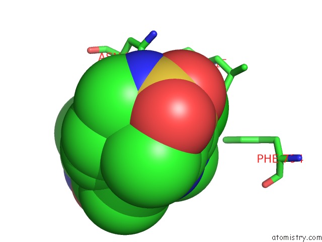
Mono view
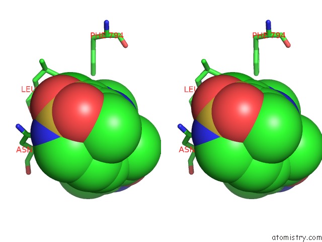
Stereo pair view
Mono view
Stereo pair view
A full contact list of Chlorine with other atoms in the Cl binding
site number 1 of Pr X-Ray Structures in Agonist Conformations Reveal Two Different Mechanisms For Partial Agonism in 11BETA-Substituted Steroids within 5.0Å range:
|
Reference:
S.J.Lusher,
H.C.Raaijmakers,
D.Vu-Pham,
B.Kazemier,
R.Bosch,
R.Mcguire,
R.Azevedo,
H.Hamersma,
K.Dechering,
A.Oubrie,
M.Van Duin,
J.De Vlieg.
X-Ray Structures of Progesterone Receptor Ligand Binding Domain in Its Agonist State Reveal Differing Mechanisms For Mixed Profiles of 11 Beta-Substituted Steroids. J. Biol. Chem. V. 287 20333 2012.
ISSN: ESSN 1083-351X
PubMed: 22535964
DOI: 10.1074/JBC.M111.308403
Page generated: Fri Jul 11 12:58:53 2025
ISSN: ESSN 1083-351X
PubMed: 22535964
DOI: 10.1074/JBC.M111.308403
Last articles
Mg in 2H03Mg in 2GZH
Mg in 2GZD
Mg in 2GWC
Mg in 2GYX
Mg in 2GYI
Mg in 2GUH
Mg in 2GWS
Mg in 2GWD
Mg in 2GSL