Atomistry » Chlorine » PDB 7lgu-7ltb » 7lkc
Atomistry »
  Chlorine »
    PDB 7lgu-7ltb »
      7lkc »

Chlorine in PDB 7lkc: Crystal Structure of Keratinimicin A

Protein crystallography data

The structure of Crystal Structure of Keratinimicin A, PDB code: 7lkc was solved by K.M.Davis, P.D.Jeffrey, M.R.Seyedsayamdost, with X-Ray Crystallography technique. A brief refinement statistics is given in the table below:

Resolution Low / High (Å) 18.97 / 0.95
Space group P 21 21 21
Cell size a, b, c (Å), α, β, γ (°) 20.72, 32.251, 37.941, 90, 90, 90
R / Rfree (%) 15.4 / 16.8

Chlorine Binding Sites:

The binding sites of Chlorine atom in the Crystal Structure of Keratinimicin A (pdb code 7lkc). This binding sites where shown within 5.0 Angstroms radius around Chlorine atom.
In total 6 binding sites of Chlorine where determined in the Crystal Structure of Keratinimicin A, PDB code: 7lkc:
Jump to Chlorine binding site number: 1; 2; 3; 4; 5; 6;

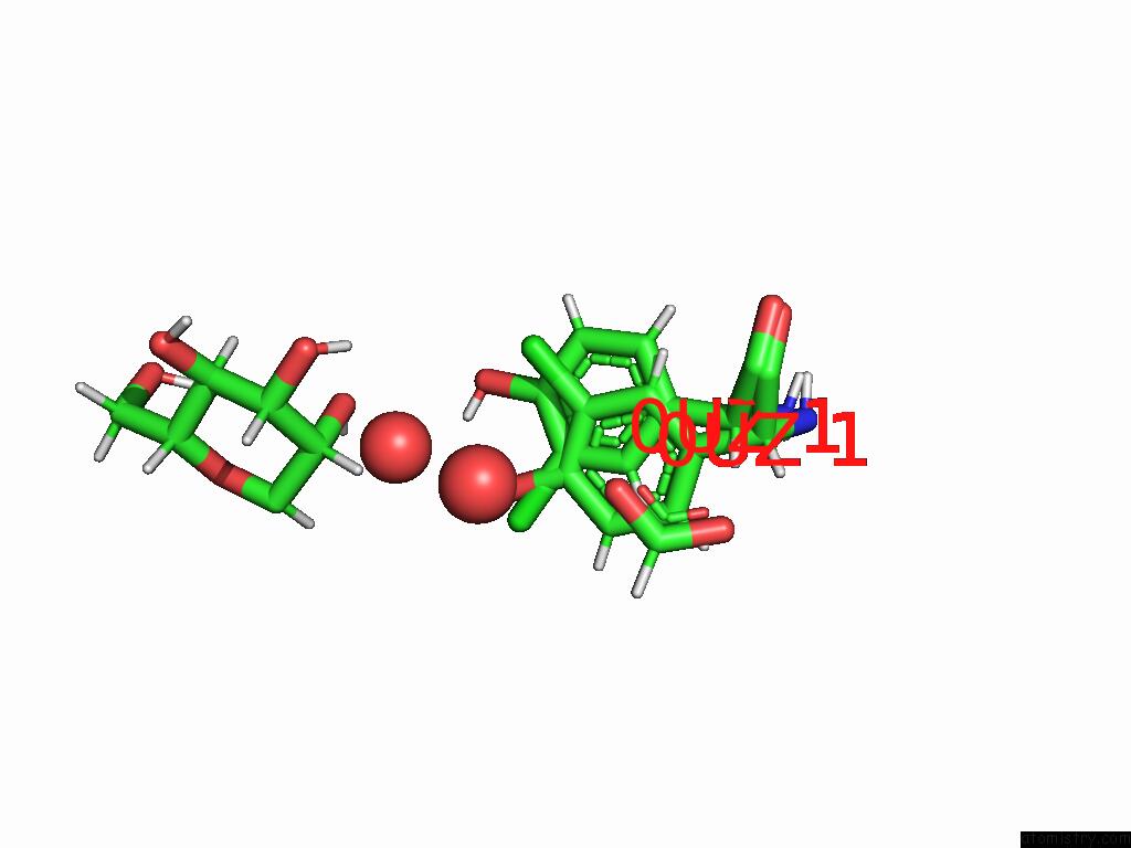
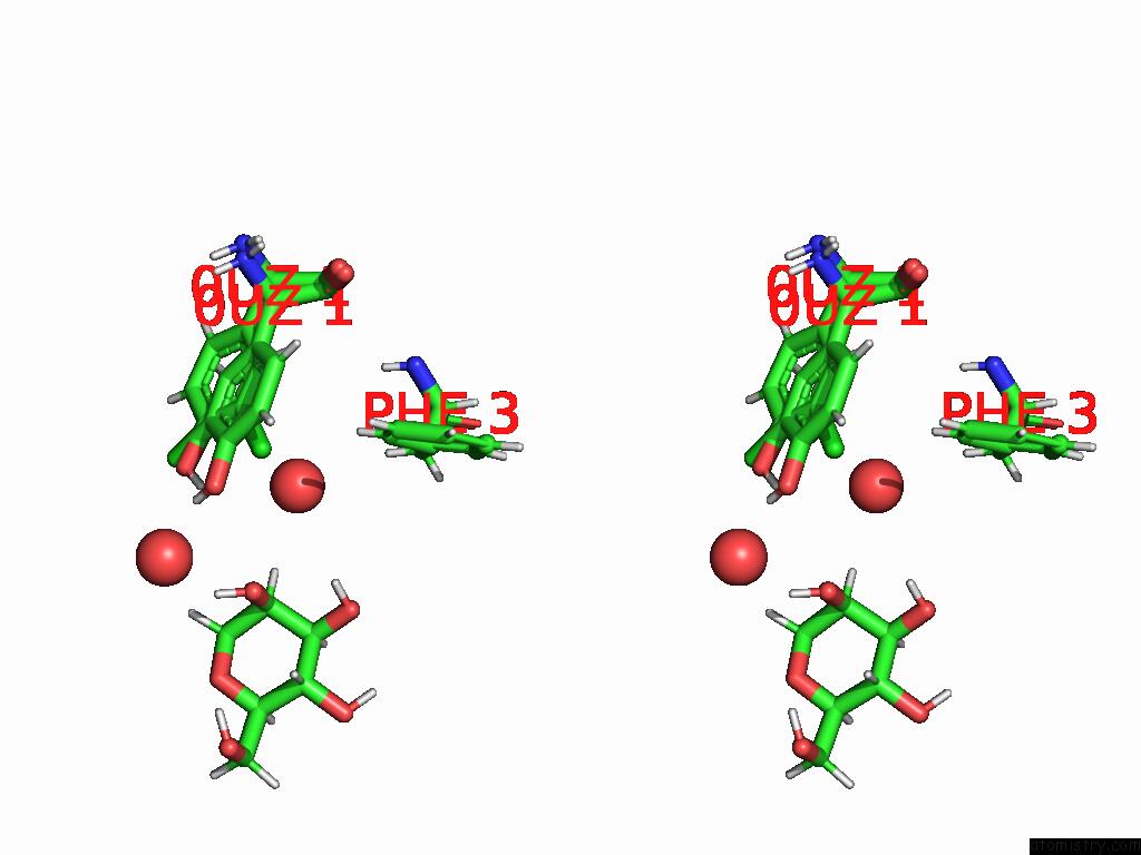
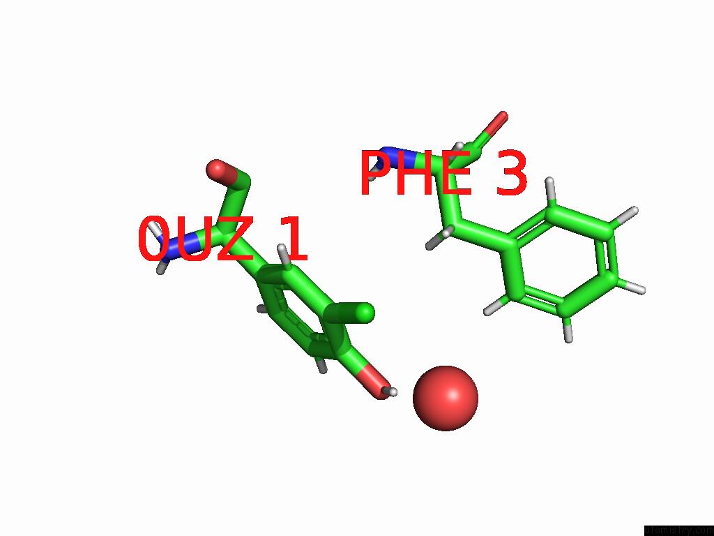
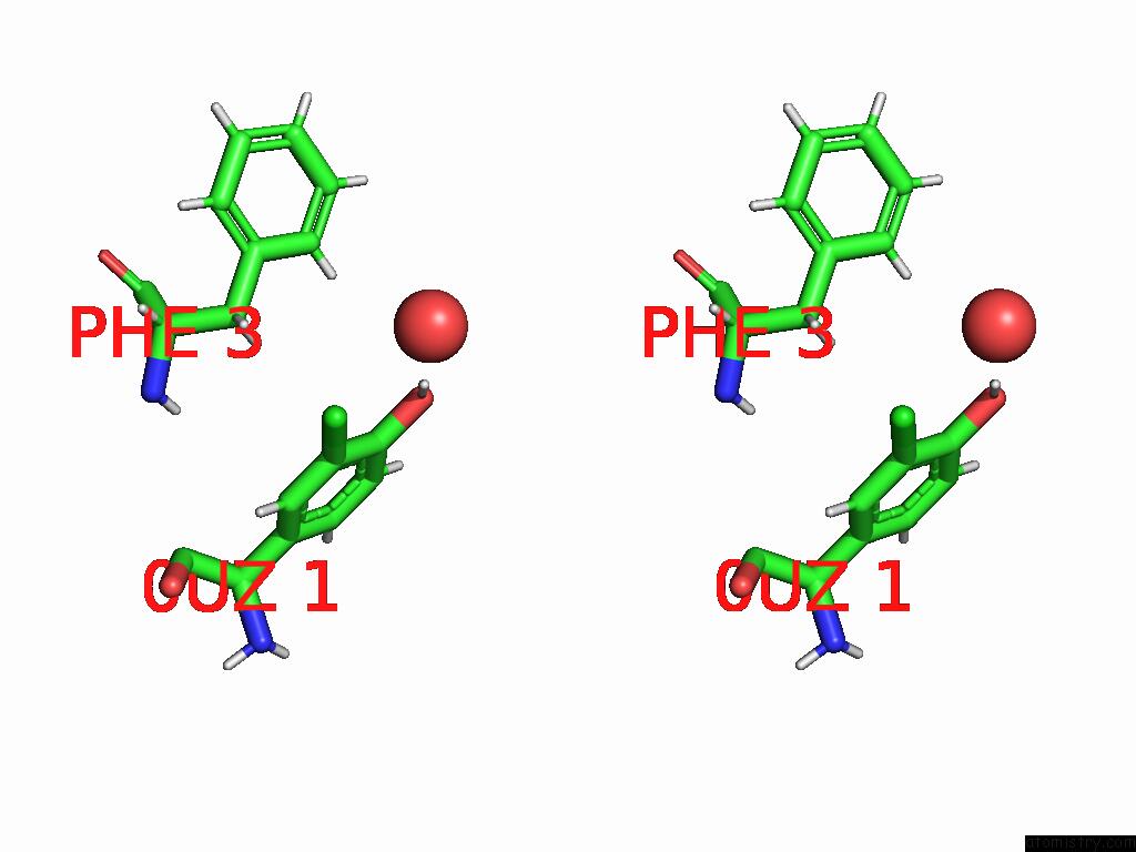
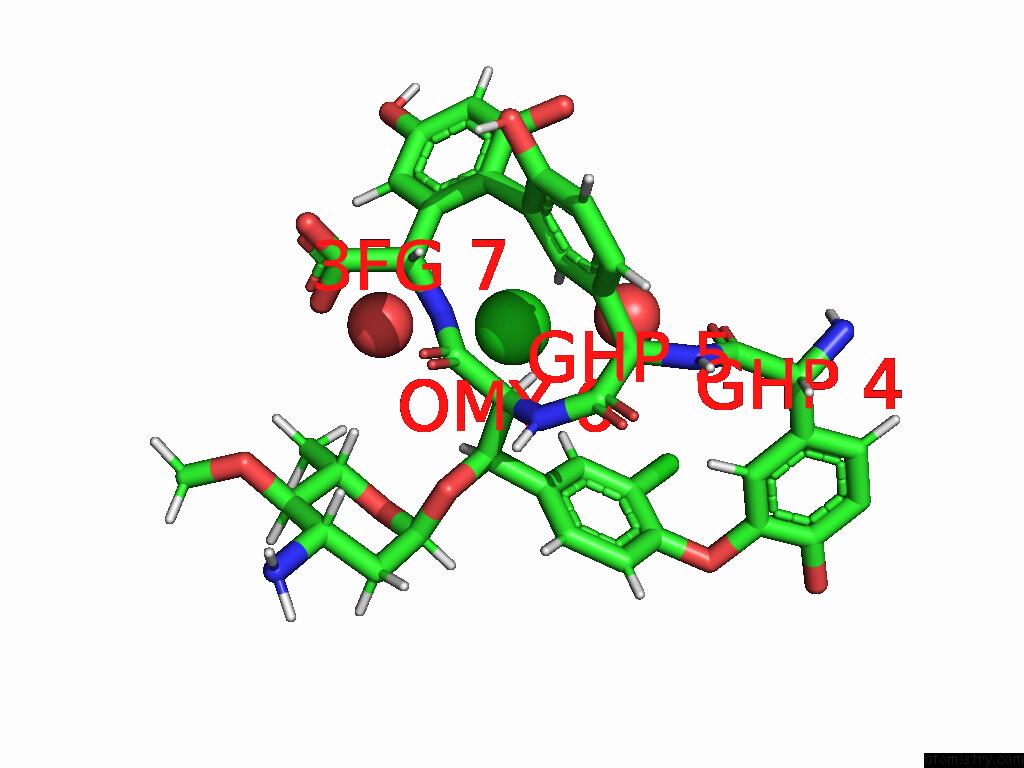
Chlorine binding site 1 out of 6 in 7lkc

Go back to Chlorine Binding Sites List in 7lkc
Chlorine binding site 1 out of 6 in the Crystal Structure of Keratinimicin A


Mono view


Stereo pair view

A full contact list of Chlorine with other atoms in the Cl binding site number 1 of Crystal Structure of Keratinimicin A within 5.0Å range:
probe atom residue distance (Å) B Occ
A:Cl1

b:10.9
occ:0.64
CL3 A:0UZ1 0.0 10.9 0.6
O4 A:0UZ1 0.7 12.1 0.4
C4 A:0UZ1 1.5 11.8 0.4
H7 A:0UZ1 1.5 14.5 0.4
C3 A:0UZ1 1.7 9.8 0.6
H8 A:0UZ1 1.9 14.4 0.4
C5 A:0UZ1 2.0 12.0 0.4
H7 A:0UZ1 2.6 12.5 0.6
C2 A:0UZ1 2.7 9.3 0.6
C4 A:0UZ1 2.7 9.8 0.6
C3 A:0UZ1 2.8 11.4 0.4
H6 A:0UZ1 2.8 11.2 0.6
O4 A:0UZ1 3.1 10.4 0.6
O A:HOH202 3.3 18.2 1.0
C6 A:0UZ1 3.4 11.9 0.4
CL3 A:0UZ1 3.6 11.2 0.4
O A:HOH209 3.7 15.2 1.0
C2 A:0UZ1 3.9 11.3 0.4
H9 A:0UZ1 4.0 14.3 0.4
C1 A:0UZ1 4.0 9.6 0.6
C5 A:0UZ1 4.0 9.8 0.6
C1 A:0UZ1 4.1 11.6 0.4
HO2 A:MAN101 4.2 22.4 1.0
H2 A:MAN101 4.3 20.9 1.0
H1 A:MAN101 4.4 18.5 1.0
O2 A:MAN101 4.5 18.7 1.0
C6 A:0UZ1 4.5 9.5 0.6
H6 A:0UZ1 4.7 13.6 0.4
H8 A:0UZ1 4.7 11.8 0.6
C2 A:MAN101 4.8 17.4 1.0
O2 A:FMT103 4.8 11.1 1.0
H A:FMT103 5.0 13.2 1.0

Chlorine binding site 2 out of 6 in 7lkc

Go back to Chlorine Binding Sites List in 7lkc
Chlorine binding site 2 out of 6 in the Crystal Structure of Keratinimicin A


Mono view


Stereo pair view

A full contact list of Chlorine with other atoms in the Cl binding site number 2 of Crystal Structure of Keratinimicin A within 5.0Å range:
probe atom residue distance (Å) B Occ
A:Cl1

b:11.2
occ:0.36
CL3 A:0UZ1 0.0 11.2 0.4
C4 A:0UZ1 1.1 9.8 0.6
C5 A:0UZ1 1.1 9.8 0.6
H8 A:0UZ1 1.3 11.8 0.6
O4 A:0UZ1 1.6 10.4 0.6
C3 A:0UZ1 1.7 11.4 0.4
H7 A:0UZ1 2.3 12.5 0.6
C6 A:0UZ1 2.3 9.5 0.6
C3 A:0UZ1 2.3 9.8 0.6
H7 A:0UZ1 2.6 14.5 0.4
C2 A:0UZ1 2.7 11.3 0.4
C4 A:0UZ1 2.7 11.8 0.4
H6 A:0UZ1 2.8 13.6 0.4
H9 A:0UZ1 3.0 11.4 0.6
O4 A:0UZ1 3.1 12.1 0.4
C2 A:0UZ1 3.1 9.3 0.6
C1 A:0UZ1 3.1 9.6 0.6
HE2 A:PHE3 3.1 13.3 1.0
HD2 A:PHE3 3.1 12.5 1.0
CL3 A:0UZ1 3.6 10.9 0.6
CE2 A:PHE3 3.7 11.1 1.0
CD2 A:PHE3 3.7 10.4 1.0
C1 A:0UZ1 4.0 11.6 0.4
H6 A:0UZ1 4.0 11.2 0.6
C5 A:0UZ1 4.0 12.0 0.4
O A:HOH202 4.0 18.2 1.0
O2 A:MAN101 4.1 18.7 1.0
O A:HOH209 4.3 15.2 1.0
HO2 A:MAN101 4.3 22.4 1.0
HO3 A:MAN101 4.5 23.4 1.0
C6 A:0UZ1 4.5 11.9 0.4
CA A:0UZ1 4.6 10.6 0.6
H8 A:0UZ1 4.7 14.4 0.4
H2 A:MAN101 4.8 20.9 1.0
CZ A:PHE3 5.0 10.8 1.0

Chlorine binding site 3 out of 6 in 7lkc

Go back to Chlorine Binding Sites List in 7lkc
Chlorine binding site 3 out of 6 in the Crystal Structure of Keratinimicin A


Mono view


Stereo pair view

A full contact list of Chlorine with other atoms in the Cl binding site number 3 of Crystal Structure of Keratinimicin A within 5.0Å range:
probe atom residue distance (Å) B Occ
A:Cl6

b:10.2
occ:1.00
CL A:OMY6 0.0 10.2 1.0
CE1 A:OMY6 1.7 8.2 1.0
HO4 G:RAM2 2.4 15.9 1.0
CD1 A:OMY6 2.7 7.8 1.0
CZ A:OMY6 2.7 7.8 1.0
HD1 A:OMY6 2.8 9.4 1.0
H62 G:RAM2 3.0 15.4 1.0
OCZ A:OMY6 3.0 7.9 1.0
C5 A:GHP4 3.2 7.8 1.0
H5 G:RAM2 3.2 14.9 1.0
O4 G:RAM2 3.4 13.3 1.0
C6 A:GHP4 3.5 7.3 1.0
H6 A:GHP4 3.6 8.8 1.0
O A:HOH207 3.7 17.1 1.0
C6 G:RAM2 3.7 12.9 1.0
C5 G:RAM2 3.9 12.4 1.0
C4 A:GHP4 4.0 8.3 1.0
H61 G:RAM2 4.0 15.4 1.0
CG A:OMY6 4.0 7.7 1.0
CE2 A:OMY6 4.0 7.8 1.0
C4 G:RAM2 4.2 13.0 1.0
O A:GHP4 4.2 7.9 1.0
H A:FMT103 4.2 13.2 1.0
H62 K:RAM2 4.3 12.7 1.0
H3 G:RAM2 4.3 16.3 1.0
C1 A:GHP4 4.3 7.5 1.0
HA A:GHP5 4.4 8.6 1.0
O4 A:GHP4 4.4 8.8 1.0
CD2 A:OMY6 4.5 7.8 1.0
H63 G:RAM2 4.7 15.4 1.0
C A:GHP4 4.7 7.4 1.0
C3 A:GHP4 4.7 8.4 1.0
HE2 A:OMY6 4.8 9.4 1.0
C3 G:RAM2 4.9 13.6 1.0
C2 A:GHP4 4.9 8.1 1.0
H61 K:RAM2 4.9 12.7 1.0
HA A:OMY6 5.0 9.1 1.0
CL A:CL104 5.0 15.0 0.7

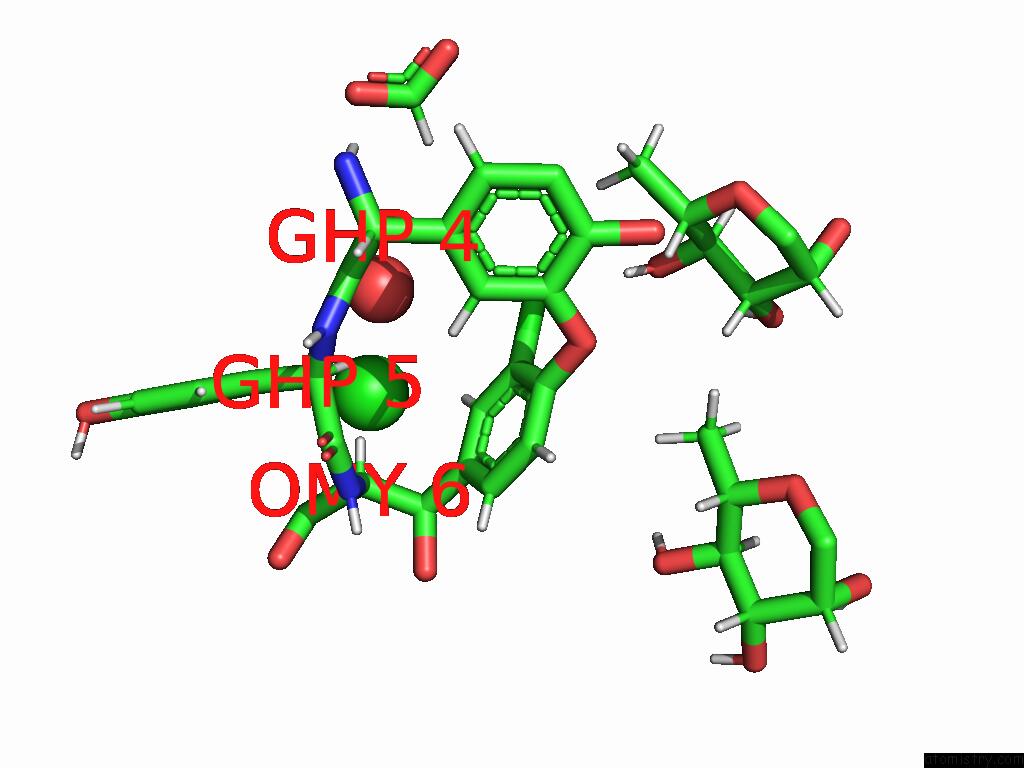
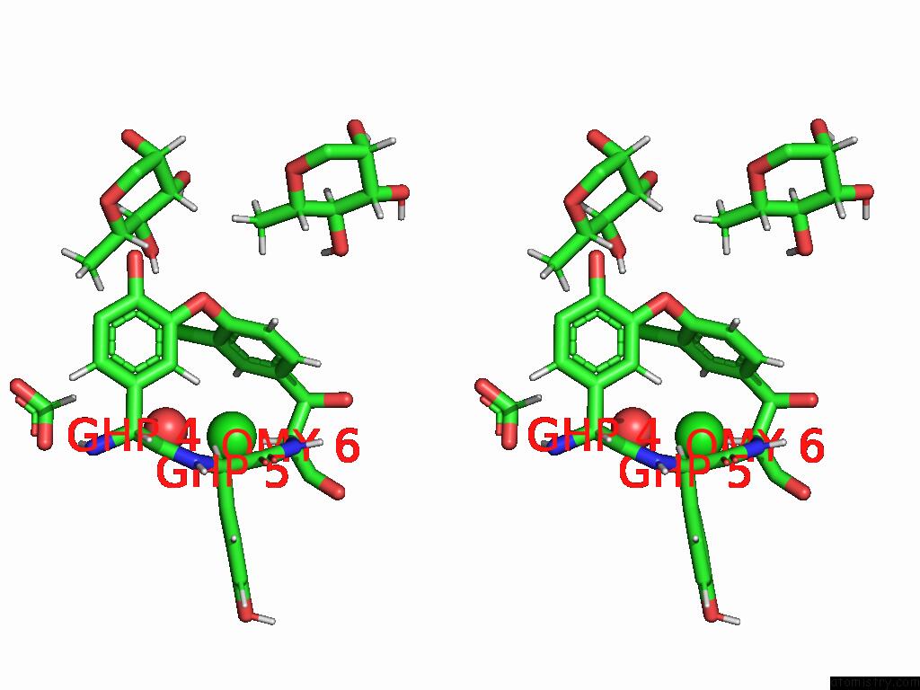
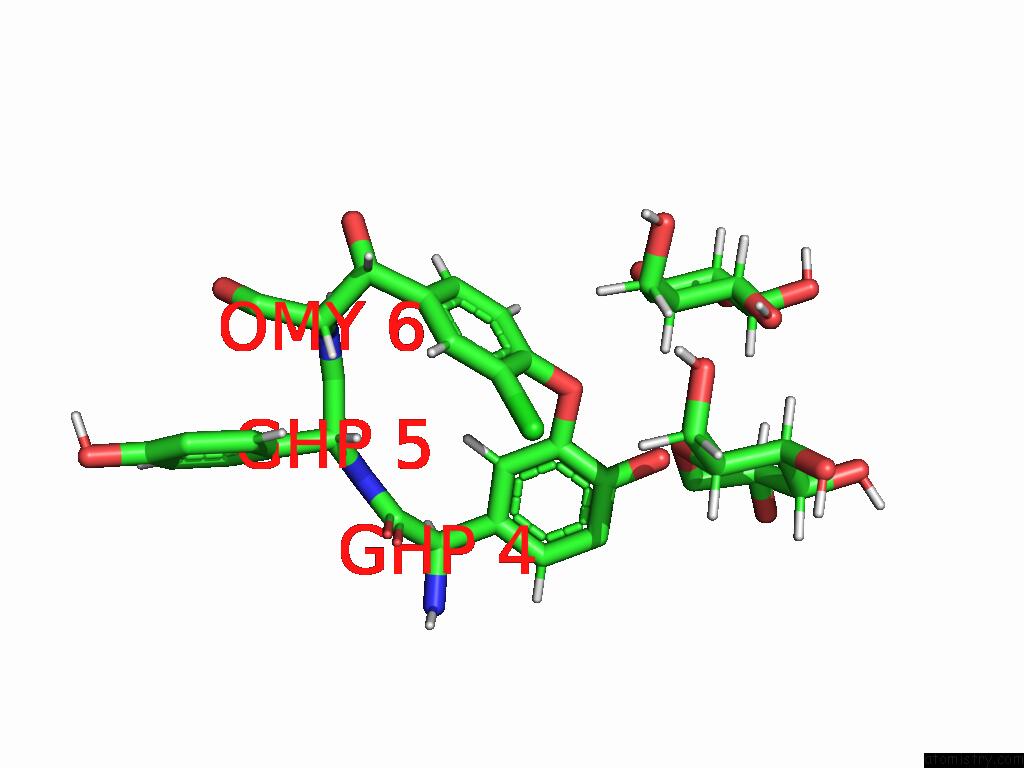
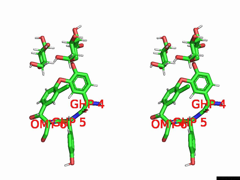
Chlorine binding site 4 out of 6 in 7lkc

Go back to Chlorine Binding Sites List in 7lkc
Chlorine binding site 4 out of 6 in the Crystal Structure of Keratinimicin A


Mono view


Stereo pair view

A full contact list of Chlorine with other atoms in the Cl binding site number 4 of Crystal Structure of Keratinimicin A within 5.0Å range:
probe atom residue distance (Å) B Occ
B:Cl1

b:18.1
occ:1.00
CL3 B:0UZ1 0.0 18.1 1.0
C3 B:0UZ1 1.7 15.7 1.0
C2 B:0UZ1 2.6 14.7 1.0
H7 B:0UZ1 2.6 19.6 1.0
C4 B:0UZ1 2.7 15.5 1.0
H6 B:0UZ1 2.7 17.6 1.0
O B:HOH211 2.8 15.9 0.7
O4 B:0UZ1 3.1 16.3 1.0
HB2 B:PHE3 3.2 15.2 1.0
C1 B:0UZ1 3.9 13.6 1.0
HB3 B:PHE3 3.9 15.2 1.0
C5 B:0UZ1 4.0 14.8 1.0
CB B:PHE3 4.0 12.7 1.0
C6 B:0UZ1 4.5 13.8 1.0
H8 B:0UZ1 4.7 17.7 1.0
H B:PHE3 4.9 12.6 1.0
N B:PHE3 4.9 10.5 1.0
CG B:PHE3 4.9 14.3 1.0
HD2 B:PHE3 4.9 19.0 1.0
CA B:PHE3 5.0 10.5 1.0

Chlorine binding site 5 out of 6 in 7lkc

Go back to Chlorine Binding Sites List in 7lkc
Chlorine binding site 5 out of 6 in the Crystal Structure of Keratinimicin A


Mono view


Stereo pair view

A full contact list of Chlorine with other atoms in the Cl binding site number 5 of Crystal Structure of Keratinimicin A within 5.0Å range:
probe atom residue distance (Å) B Occ
B:Cl6

b:11.4
occ:1.00
CL B:OMY6 0.0 11.4 1.0
CE1 B:OMY6 1.7 9.2 1.0
CD1 B:OMY6 2.7 8.9 1.0
CZ B:OMY6 2.7 8.5 1.0
HD1 B:OMY6 2.8 10.7 1.0
H62 K:BGC1 2.9 14.6 1.0
OCZ B:OMY6 3.0 8.6 1.0
C5 B:GHP4 3.2 8.0 1.0
C6 B:GHP4 3.5 8.2 1.0
H6 B:GHP4 3.5 9.9 1.0
O5 K:BGC1 3.6 9.9 1.0
H62 G:BGC1 3.7 15.0 1.0
C6 K:BGC1 3.8 12.1 1.0
C4 B:GHP4 3.9 8.2 1.0
CE2 B:OMY6 4.0 8.4 1.0
CG B:OMY6 4.0 8.5 1.0
H61 G:BGC1 4.0 15.0 1.0
HO6 K:BGC1 4.1 15.7 1.0
O6 K:BGC1 4.1 13.1 1.0
C5 K:BGC1 4.2 10.8 1.0
HA B:GHP5 4.3 9.4 1.0
C1 B:GHP4 4.3 8.0 1.0
O4 B:GHP4 4.4 8.7 1.0
O B:GHP4 4.4 8.7 1.0
C6 G:BGC1 4.4 12.5 1.0
H5 K:BGC1 4.4 13.0 1.0
C1 K:BGC1 4.4 9.3 1.0
CD2 B:OMY6 4.5 8.2 1.0
H5 G:BGC1 4.5 12.9 1.0
H61 K:BGC1 4.6 14.6 1.0
C3 B:GHP4 4.7 8.3 1.0
HE2 B:OMY6 4.7 10.1 1.0
C B:GHP4 4.8 7.9 1.0
HA B:OMY6 4.8 10.1 1.0
C2 B:GHP4 4.9 8.2 1.0

Chlorine binding site 6 out of 6 in 7lkc

Go back to Chlorine Binding Sites List in 7lkc
Chlorine binding site 6 out of 6 in the Crystal Structure of Keratinimicin A


Mono view


Stereo pair view

A full contact list of Chlorine with other atoms in the Cl binding site number 6 of Crystal Structure of Keratinimicin A within 5.0Å range:
probe atom residue distance (Å) B Occ
A:Cl104

b:15.0
occ:0.66
HN1 A:3FG7 2.2 10.2 1.0
HC2 A:GHP5 2.5 8.9 1.0
HA A:OMY6 2.6 9.1 1.0
O A:HOH207 2.7 17.1 1.0
HD1 A:OMY6 2.8 9.4 1.0
N A:3FG7 3.0 8.5 1.0
O A:HOH211 3.2 23.4 0.9
HB A:OMY6 3.3 9.1 1.0
CA A:OMY6 3.4 7.6 1.0
HA A:GHP5 3.5 8.6 1.0
C2 A:GHP5 3.6 7.4 1.0
CD1 A:OMY6 3.7 7.8 1.0
C A:OMY6 3.7 8.5 1.0
CB A:OMY6 3.7 7.6 1.0
CB A:3FG7 3.9 9.1 1.0
CA A:3FG7 4.0 9.2 1.0
CG1 A:3FG7 4.1 8.9 1.0
CG A:OMY6 4.2 7.7 1.0
C3 A:GHP5 4.3 8.3 1.0
CG2 A:3FG7 4.3 9.9 1.0
CA A:GHP5 4.5 7.2 1.0
C1 A:GHP5 4.5 7.4 1.0
HG2 A:3FG7 4.7 11.9 1.0
N A:OMY6 4.7 7.2 1.0
H6 A:YBJ102 4.7 12.9 1.0
CE1 A:OMY6 4.7 8.2 1.0
HA A:3FG7 4.7 11.1 1.0
O A:3FG7 4.8 11.7 1.0
CD1 A:3FG7 4.8 10.0 1.0
H A:YBJ102 4.8 11.5 1.0
O A:OMY6 4.9 9.6 1.0
C A:3FG7 4.9 10.7 1.0
ODE A:OMY6 4.9 7.7 1.0
CD2 A:3FG7 5.0 10.6 1.0
CL A:OMY6 5.0 10.2 1.0
O A:GHP4 5.0 7.9 1.0

Reference:

K.L.Mcwhorter, V.T.Chioti, F.Xu, P.D.Jeffrey, K.M.Davis, M.R.Seyedsayamdost. Structural and Functional Analysis of Keratinicyclin Reveals Synergistic Antibiosis with Vancomycin Against Clostridium Difficile To Be Published.
Page generated: Sun Jul 13 03:46:01 2025

Last articles

Mg in 4X5C
Mg in 4X5E
Mg in 4X5V
Mg in 4X5B
Mg in 4X59
Mg in 4X58
Mg in 4X4V
Mg in 4X4S
Mg in 4X4R
Mg in 4X4Q
© Copyright 2008-2020 by atomistry.com
Home   |    Site Map   |    Copyright   |    Contact us   |    Privacy