Chlorine »
PDB 2pgc-2px2 »
2pvv »
Chlorine in PDB 2pvv: Structure of Human Glutamate Carboxypeptidase II (Gcpii) in Complex with L-Serine-O-Sulfate
Enzymatic activity of Structure of Human Glutamate Carboxypeptidase II (Gcpii) in Complex with L-Serine-O-Sulfate
All present enzymatic activity of Structure of Human Glutamate Carboxypeptidase II (Gcpii) in Complex with L-Serine-O-Sulfate:
3.4.17.21;
3.4.17.21;
Protein crystallography data
The structure of Structure of Human Glutamate Carboxypeptidase II (Gcpii) in Complex with L-Serine-O-Sulfate, PDB code: 2pvv
was solved by
C.Barinka,
J.Lubkowski,
with X-Ray Crystallography technique. A brief refinement statistics is given in the table below:
| Resolution Low / High (Å) | 30.00 / 2.11 |
| Space group | I 2 2 2 |
| Cell size a, b, c (Å), α, β, γ (°) | 101.617, 130.220, 158.975, 90.00, 90.00, 90.00 |
| R / Rfree (%) | 17.4 / 21.2 |
Other elements in 2pvv:
The structure of Structure of Human Glutamate Carboxypeptidase II (Gcpii) in Complex with L-Serine-O-Sulfate also contains other interesting chemical elements:
| Calcium | (Ca) | 1 atom |
| Zinc | (Zn) | 2 atoms |
Chlorine Binding Sites:
The binding sites of Chlorine atom in the Structure of Human Glutamate Carboxypeptidase II (Gcpii) in Complex with L-Serine-O-Sulfate
(pdb code 2pvv). This binding sites where shown within
5.0 Angstroms radius around Chlorine atom.
In total only one binding site of Chlorine was determined in the Structure of Human Glutamate Carboxypeptidase II (Gcpii) in Complex with L-Serine-O-Sulfate, PDB code: 2pvv:
In total only one binding site of Chlorine was determined in the Structure of Human Glutamate Carboxypeptidase II (Gcpii) in Complex with L-Serine-O-Sulfate, PDB code: 2pvv:
Chlorine binding site 1 out of 1 in 2pvv
Go back to
Chlorine binding site 1 out
of 1 in the Structure of Human Glutamate Carboxypeptidase II (Gcpii) in Complex with L-Serine-O-Sulfate
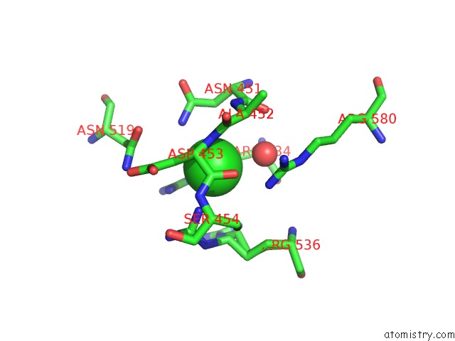
Mono view
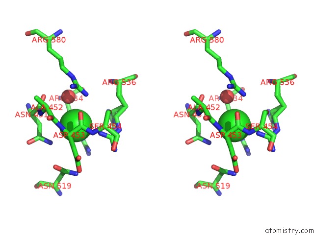
Stereo pair view
Mono view
Stereo pair view
A full contact list of Chlorine with other atoms in the Cl binding
site number 1 of Structure of Human Glutamate Carboxypeptidase II (Gcpii) in Complex with L-Serine-O-Sulfate within 5.0Å range:
|
Reference:
C.Barinka,
M.Rovenska,
P.Mlcochova,
K.Hlouchova,
A.Plechanovova,
P.Majer,
T.Tsukamoto,
B.S.Slusher,
J.Konvalinka,
J.Lubkowski.
Structural Insight Into the Pharmacophore Pocket of Human Glutamate Carboxypeptidase II. J.Med.Chem. V. 50 3267 2007.
ISSN: ISSN 0022-2623
PubMed: 17567119
DOI: 10.1021/JM070133W
Page generated: Thu Jul 10 23:56:08 2025
ISSN: ISSN 0022-2623
PubMed: 17567119
DOI: 10.1021/JM070133W
Last articles
Cl in 3UHOCl in 3UIA
Cl in 3UGV
Cl in 3UHF
Cl in 3UGR
Cl in 3UHA
Cl in 3UG8
Cl in 3UFT
Cl in 3UFI
Cl in 3UG2