Chlorine »
PDB 3rv4-3s8n »
3s4k »
Chlorine in PDB 3s4k: Structure of A Putative Esterase RV1847/MT1895 From Mycobacterium Tuberculosis
Protein crystallography data
The structure of Structure of A Putative Esterase RV1847/MT1895 From Mycobacterium Tuberculosis, PDB code: 3s4k
was solved by
Seattle Structural Genomics Center For Infectious Disease (Ssgcid),
with X-Ray Crystallography technique. A brief refinement statistics is given in the table below:
| Resolution Low / High (Å) | 38.82 / 1.70 |
| Space group | P 1 2 1 |
| Cell size a, b, c (Å), α, β, γ (°) | 41.722, 50.642, 60.506, 90.00, 92.76, 90.00 |
| R / Rfree (%) | 17.1 / 19.7 |
Chlorine Binding Sites:
The binding sites of Chlorine atom in the Structure of A Putative Esterase RV1847/MT1895 From Mycobacterium Tuberculosis
(pdb code 3s4k). This binding sites where shown within
5.0 Angstroms radius around Chlorine atom.
In total 4 binding sites of Chlorine where determined in the Structure of A Putative Esterase RV1847/MT1895 From Mycobacterium Tuberculosis, PDB code: 3s4k:
Jump to Chlorine binding site number: 1; 2; 3; 4;
In total 4 binding sites of Chlorine where determined in the Structure of A Putative Esterase RV1847/MT1895 From Mycobacterium Tuberculosis, PDB code: 3s4k:
Jump to Chlorine binding site number: 1; 2; 3; 4;
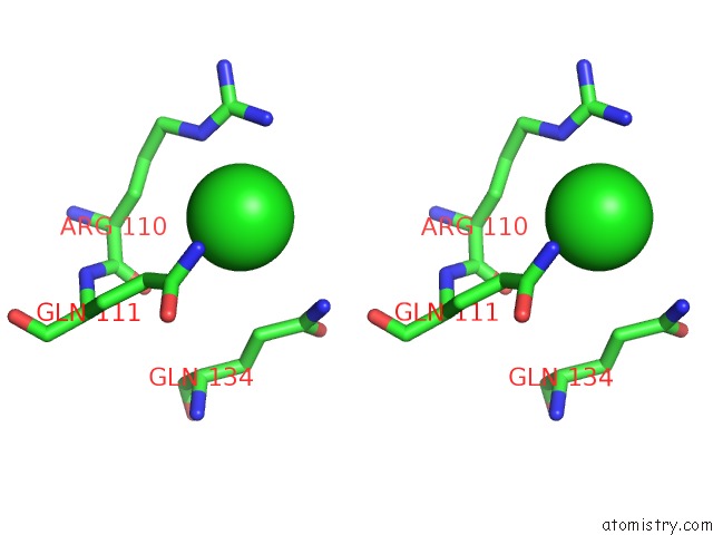
Chlorine binding site 1 out of 4 in 3s4k
Go back to
Chlorine binding site 1 out
of 4 in the Structure of A Putative Esterase RV1847/MT1895 From Mycobacterium Tuberculosis

Mono view
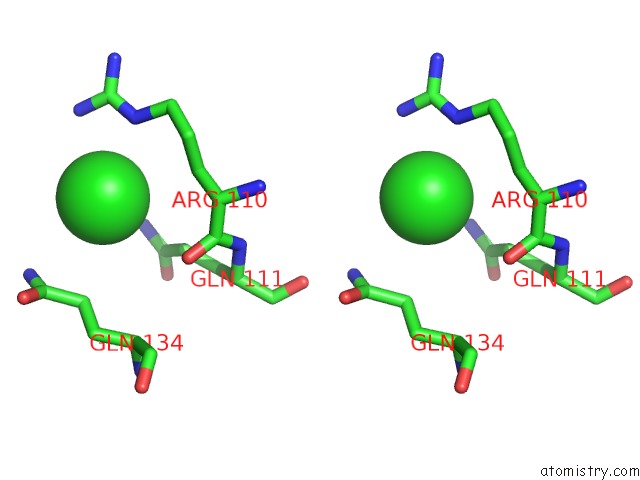
Stereo pair view
Mono view
Stereo pair view
A full contact list of Chlorine with other atoms in the Cl binding
site number 1 of Structure of A Putative Esterase RV1847/MT1895 From Mycobacterium Tuberculosis within 5.0Å range:
|
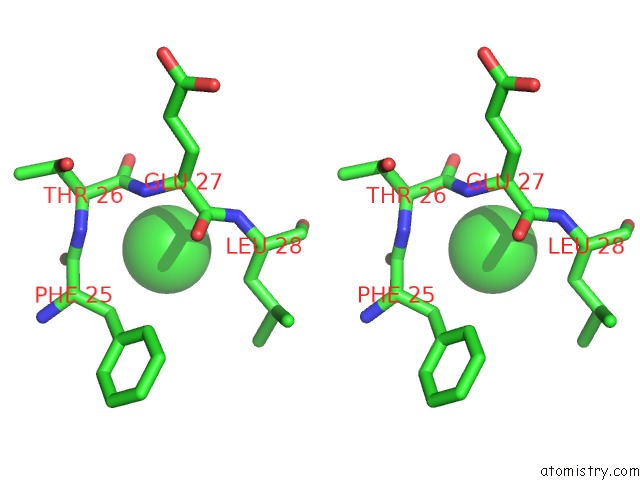
Chlorine binding site 2 out of 4 in 3s4k
Go back to
Chlorine binding site 2 out
of 4 in the Structure of A Putative Esterase RV1847/MT1895 From Mycobacterium Tuberculosis
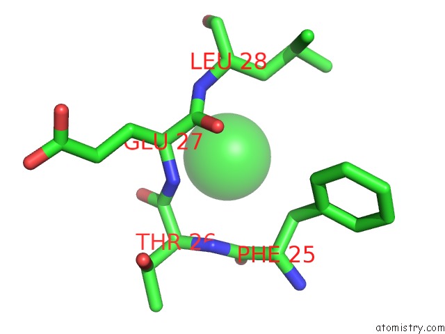
Mono view

Stereo pair view
Mono view
Stereo pair view
A full contact list of Chlorine with other atoms in the Cl binding
site number 2 of Structure of A Putative Esterase RV1847/MT1895 From Mycobacterium Tuberculosis within 5.0Å range:
|
Chlorine binding site 3 out of 4 in 3s4k
Go back to
Chlorine binding site 3 out
of 4 in the Structure of A Putative Esterase RV1847/MT1895 From Mycobacterium Tuberculosis

Mono view
Stereo pair view
Mono view
Stereo pair view
A full contact list of Chlorine with other atoms in the Cl binding
site number 3 of Structure of A Putative Esterase RV1847/MT1895 From Mycobacterium Tuberculosis within 5.0Å range:
|
Chlorine binding site 4 out of 4 in 3s4k
Go back to
Chlorine binding site 4 out
of 4 in the Structure of A Putative Esterase RV1847/MT1895 From Mycobacterium Tuberculosis

Mono view
Stereo pair view
Mono view
Stereo pair view
A full contact list of Chlorine with other atoms in the Cl binding
site number 4 of Structure of A Putative Esterase RV1847/MT1895 From Mycobacterium Tuberculosis within 5.0Å range:
|
Reference:
M.C.Clifton,
T.E.Edwards,
B.Sankaran,
Seattle Structural Genomics Center For Infectious Disease(Ssgcid).
Structure of A Putative Esterase RV1847/MT1895 From Mycobacterium Tuberculosis To Be Published.
Page generated: Fri Jul 11 10:06:04 2025
Last articles
Cl in 4H1TCl in 4H1N
Cl in 4GZE
Cl in 4H16
Cl in 4GYF
Cl in 4H12
Cl in 4GYZ
Cl in 4GZD
Cl in 4GYO
Cl in 4GZC