Chlorine »
PDB 3ue8-3unh »
3ui4 »
Chlorine in PDB 3ui4: 0.8 A Resolution Crystal Structure of Human Parvulin 14
Enzymatic activity of 0.8 A Resolution Crystal Structure of Human Parvulin 14
All present enzymatic activity of 0.8 A Resolution Crystal Structure of Human Parvulin 14:
5.2.1.8;
5.2.1.8;
Protein crystallography data
The structure of 0.8 A Resolution Crystal Structure of Human Parvulin 14, PDB code: 3ui4
was solved by
J.W.Mueller,
N.M.Link,
A.Matena,
L.Hoppstock,
A.Rueppel,
P.Bayer,
W.Blankenfeldt,
with X-Ray Crystallography technique. A brief refinement statistics is given in the table below:
| Resolution Low / High (Å) | 34.80 / 0.80 |
| Space group | P 21 21 21 |
| Cell size a, b, c (Å), α, β, γ (°) | 34.043, 46.966, 51.826, 90.00, 90.00, 90.00 |
| R / Rfree (%) | 9.5 / 9.8 |
Chlorine Binding Sites:
The binding sites of Chlorine atom in the 0.8 A Resolution Crystal Structure of Human Parvulin 14
(pdb code 3ui4). This binding sites where shown within
5.0 Angstroms radius around Chlorine atom.
In total 2 binding sites of Chlorine where determined in the 0.8 A Resolution Crystal Structure of Human Parvulin 14, PDB code: 3ui4:
Jump to Chlorine binding site number: 1; 2;
In total 2 binding sites of Chlorine where determined in the 0.8 A Resolution Crystal Structure of Human Parvulin 14, PDB code: 3ui4:
Jump to Chlorine binding site number: 1; 2;
Chlorine binding site 1 out of 2 in 3ui4
Go back to
Chlorine binding site 1 out
of 2 in the 0.8 A Resolution Crystal Structure of Human Parvulin 14
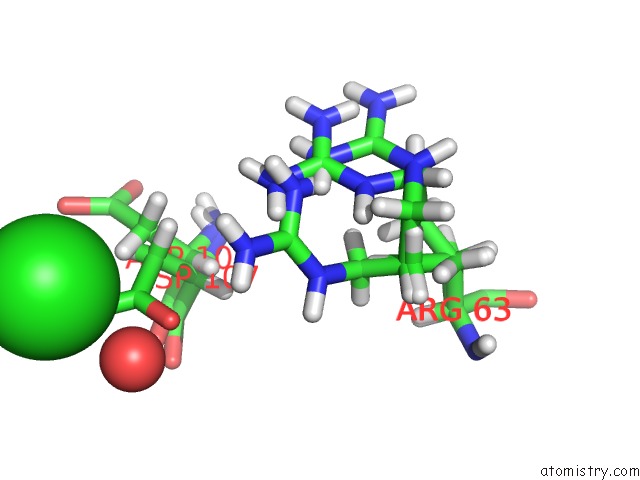
Mono view
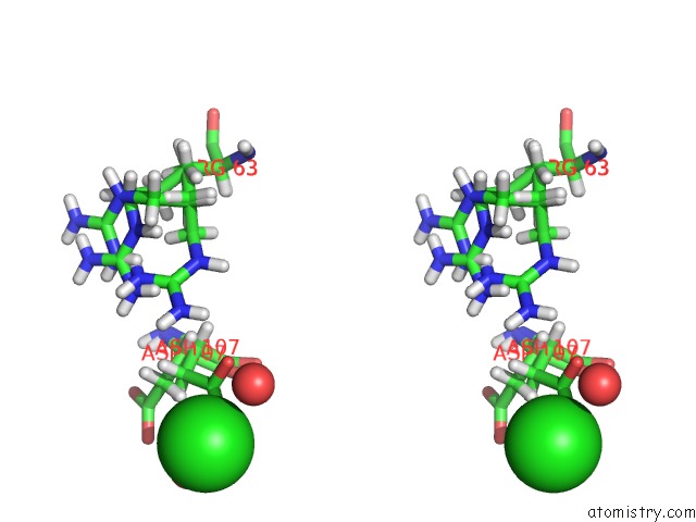
Stereo pair view
Mono view
Stereo pair view
A full contact list of Chlorine with other atoms in the Cl binding
site number 1 of 0.8 A Resolution Crystal Structure of Human Parvulin 14 within 5.0Å range:
|
Chlorine binding site 2 out of 2 in 3ui4
Go back to
Chlorine binding site 2 out
of 2 in the 0.8 A Resolution Crystal Structure of Human Parvulin 14
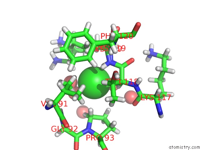
Mono view
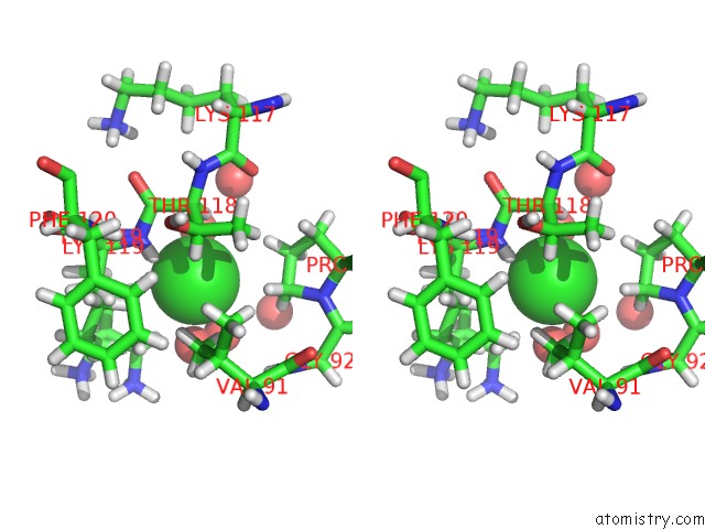
Stereo pair view
Mono view
Stereo pair view
A full contact list of Chlorine with other atoms in the Cl binding
site number 2 of 0.8 A Resolution Crystal Structure of Human Parvulin 14 within 5.0Å range:
|
Reference:
J.W.Mueller,
N.M.Link,
A.Matena,
L.Hoppstock,
A.Ruppel,
P.Bayer,
W.Blankenfeldt.
Crystallographic Proof For An Extended Hydrogen-Bonding Network in Small Prolyl Isomerases. J.Am.Chem.Soc. V. 133 20096 2011.
ISSN: ISSN 0002-7863
PubMed: 22081960
DOI: 10.1021/JA2086195
Page generated: Fri Jul 11 11:16:07 2025
ISSN: ISSN 0002-7863
PubMed: 22081960
DOI: 10.1021/JA2086195
Last articles
Cl in 5UN3Cl in 5UOA
Cl in 5UNN
Cl in 5UMH
Cl in 5UMM
Cl in 5UM6
Cl in 5UM1
Cl in 5ULX
Cl in 5ULT
Cl in 5UKA