Chlorine »
PDB 4q69-4qgp »
4q7z »
Chlorine in PDB 4q7z: Neutrophil Serine Protease 4 (PRSS57) with Phe-Phe-Arg- Chloromethylketone (Ffr-Cmk)
Protein crystallography data
The structure of Neutrophil Serine Protease 4 (PRSS57) with Phe-Phe-Arg- Chloromethylketone (Ffr-Cmk), PDB code: 4q7z
was solved by
C.Eigenbrot,
S.J.Lin,
K.C.Dong,
with X-Ray Crystallography technique. A brief refinement statistics is given in the table below:
| Resolution Low / High (Å) | 46.91 / 1.40 |
| Space group | P 21 21 21 |
| Cell size a, b, c (Å), α, β, γ (°) | 54.986, 64.466, 68.383, 90.00, 90.00, 90.00 |
| R / Rfree (%) | 19.2 / 21.5 |
Chlorine Binding Sites:
The binding sites of Chlorine atom in the Neutrophil Serine Protease 4 (PRSS57) with Phe-Phe-Arg- Chloromethylketone (Ffr-Cmk)
(pdb code 4q7z). This binding sites where shown within
5.0 Angstroms radius around Chlorine atom.
In total only one binding site of Chlorine was determined in the Neutrophil Serine Protease 4 (PRSS57) with Phe-Phe-Arg- Chloromethylketone (Ffr-Cmk), PDB code: 4q7z:
In total only one binding site of Chlorine was determined in the Neutrophil Serine Protease 4 (PRSS57) with Phe-Phe-Arg- Chloromethylketone (Ffr-Cmk), PDB code: 4q7z:
Chlorine binding site 1 out of 1 in 4q7z
Go back to
Chlorine binding site 1 out
of 1 in the Neutrophil Serine Protease 4 (PRSS57) with Phe-Phe-Arg- Chloromethylketone (Ffr-Cmk)
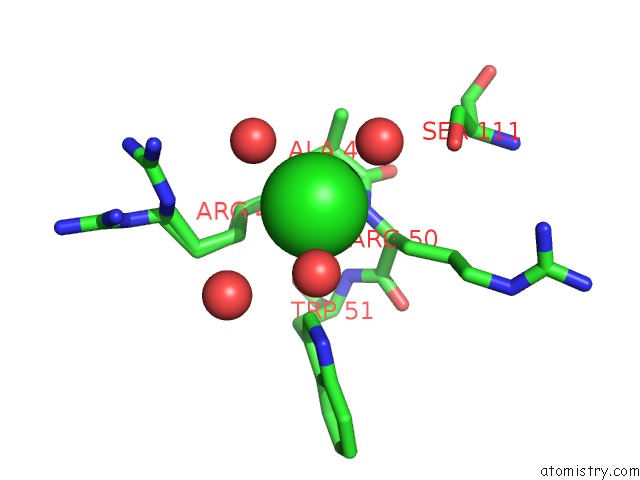
Mono view
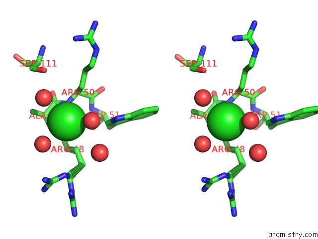
Stereo pair view
Mono view
Stereo pair view
A full contact list of Chlorine with other atoms in the Cl binding
site number 1 of Neutrophil Serine Protease 4 (PRSS57) with Phe-Phe-Arg- Chloromethylketone (Ffr-Cmk) within 5.0Å range:
|
Reference:
S.J.Lin,
K.C.Dong,
C.Eigenbrot,
M.Van Lookeren Campagne,
D.Kirchhofer.
Structures of Neutrophil Serine Protease 4 Reveal An Unusual Mechanism of Substrate Recognition By A Trypsin-Fold Protease. Structure V. 22 1333 2014.
ISSN: ISSN 0969-2126
PubMed: 25156428
DOI: 10.1016/J.STR.2014.07.008
Page generated: Fri Jul 11 20:42:44 2025
ISSN: ISSN 0969-2126
PubMed: 25156428
DOI: 10.1016/J.STR.2014.07.008
Last articles
Cl in 5DNICl in 5DN9
Cl in 5DN5
Cl in 5DLA
Cl in 5DN4
Cl in 5DM9
Cl in 5DML
Cl in 5DLX
Cl in 5DLO
Cl in 5DKJ