Chlorine »
PDB 5eaf-5eic »
5ec4 »
Chlorine in PDB 5ec4: Crystal Structure of Acetyltransferase Eis From Mycobacterium Tuberculosis in Complex with Inhibitor 13G and Coa
Protein crystallography data
The structure of Crystal Structure of Acetyltransferase Eis From Mycobacterium Tuberculosis in Complex with Inhibitor 13G and Coa, PDB code: 5ec4
was solved by
C.S.Gajadeera,
C.Hou,
S.Garneau-Tsodikova,
O.V.Tsodikov,
with X-Ray Crystallography technique. A brief refinement statistics is given in the table below:
| Resolution Low / High (Å) | 40.00 / 2.21 |
| Space group | H 3 2 |
| Cell size a, b, c (Å), α, β, γ (°) | 175.632, 175.632, 122.215, 90.00, 90.00, 120.00 |
| R / Rfree (%) | 19.8 / 23.8 |
Chlorine Binding Sites:
The binding sites of Chlorine atom in the Crystal Structure of Acetyltransferase Eis From Mycobacterium Tuberculosis in Complex with Inhibitor 13G and Coa
(pdb code 5ec4). This binding sites where shown within
5.0 Angstroms radius around Chlorine atom.
In total only one binding site of Chlorine was determined in the Crystal Structure of Acetyltransferase Eis From Mycobacterium Tuberculosis in Complex with Inhibitor 13G and Coa, PDB code: 5ec4:
In total only one binding site of Chlorine was determined in the Crystal Structure of Acetyltransferase Eis From Mycobacterium Tuberculosis in Complex with Inhibitor 13G and Coa, PDB code: 5ec4:
Chlorine binding site 1 out of 1 in 5ec4
Go back to
Chlorine binding site 1 out
of 1 in the Crystal Structure of Acetyltransferase Eis From Mycobacterium Tuberculosis in Complex with Inhibitor 13G and Coa
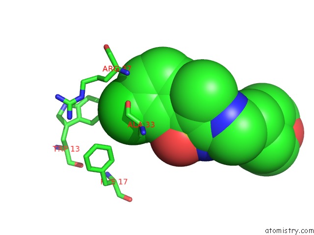
Mono view
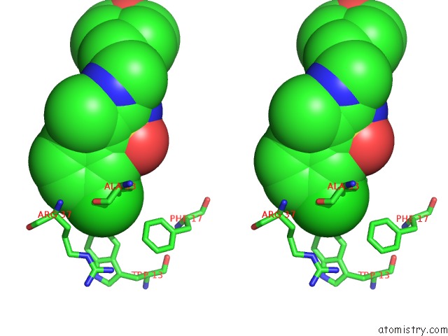
Stereo pair view
Mono view
Stereo pair view
A full contact list of Chlorine with other atoms in the Cl binding
site number 1 of Crystal Structure of Acetyltransferase Eis From Mycobacterium Tuberculosis in Complex with Inhibitor 13G and Coa within 5.0Å range:
|
Reference:
M.J.Willby,
K.D.Green,
C.S.Gajadeera,
C.Hou,
O.V.Tsodikov,
J.E.Posey,
S.Garneau-Tsodikova.
Potent Inhibitors of Acetyltransferase Eis Overcome Kanamycin Resistance in Mycobacterium Tuberculosis. Acs Chem.Biol. V. 11 1639 2016.
ISSN: ESSN 1554-8937
PubMed: 27010218
DOI: 10.1021/ACSCHEMBIO.6B00110
Page generated: Sat Jul 12 01:37:48 2025
ISSN: ESSN 1554-8937
PubMed: 27010218
DOI: 10.1021/ACSCHEMBIO.6B00110
Last articles
Cl in 5NAECl in 5NAB
Cl in 5N9P
Cl in 5NA7
Cl in 5NA5
Cl in 5N9L
Cl in 5N9N
Cl in 5N9C
Cl in 5N9K
Cl in 5N91