Chlorine in PDB 5g3s: The Structure of the L-Tryptophan Oxidase Vioa From Chromobacterium Violaceum - Samarium Derivative
Enzymatic activity of The Structure of the L-Tryptophan Oxidase Vioa From Chromobacterium Violaceum - Samarium Derivative
All present enzymatic activity of The Structure of the L-Tryptophan Oxidase Vioa From Chromobacterium Violaceum - Samarium Derivative:
1.4.3.23;
Protein crystallography data
The structure of The Structure of the L-Tryptophan Oxidase Vioa From Chromobacterium Violaceum - Samarium Derivative, PDB code: 5g3s
was solved by
J.Krausze,
J.Rabe,
J.Moser,
with X-Ray Crystallography technique. A brief refinement statistics is given in the table below:
Resolution Low / High (Å)
|
19.98 /
2.08
|
Space group
|
P 1 21 1
|
Cell size a, b, c (Å), α, β, γ (°)
|
67.880,
87.070,
78.020,
90.00,
112.95,
90.00
|
R / Rfree (%)
|
16.5 /
18.8
|
Other elements in 5g3s:
The structure of The Structure of the L-Tryptophan Oxidase Vioa From Chromobacterium Violaceum - Samarium Derivative also contains other interesting chemical elements:
Chlorine Binding Sites:
The binding sites of Chlorine atom in the The Structure of the L-Tryptophan Oxidase Vioa From Chromobacterium Violaceum - Samarium Derivative
(pdb code 5g3s). This binding sites where shown within
5.0 Angstroms radius around Chlorine atom.
In total 3 binding sites of Chlorine where determined in the
The Structure of the L-Tryptophan Oxidase Vioa From Chromobacterium Violaceum - Samarium Derivative, PDB code: 5g3s:
Jump to Chlorine binding site number:
1;
2;
3;
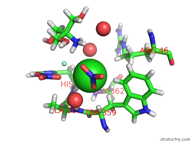
Chlorine binding site 1 out
of 3 in 5g3s
Go back to
Chlorine Binding Sites List in 5g3s
Chlorine binding site 1 out
of 3 in the The Structure of the L-Tryptophan Oxidase Vioa From Chromobacterium Violaceum - Samarium Derivative
Mono view
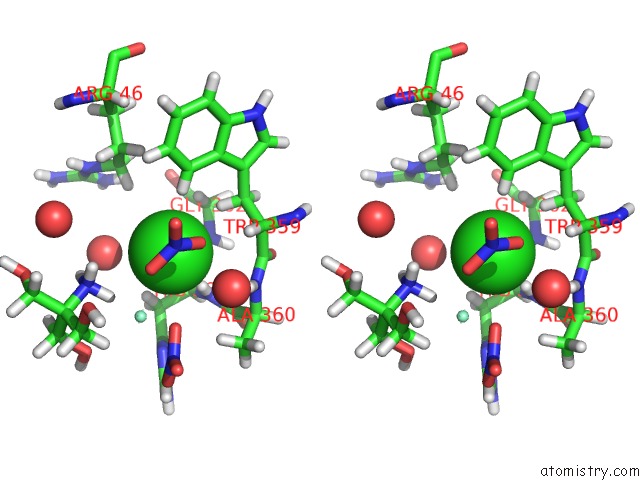
Stereo pair view
|
A full contact list of Chlorine with other atoms in the Cl binding
site number 1 of The Structure of the L-Tryptophan Oxidase Vioa From Chromobacterium Violaceum - Samarium Derivative within 5.0Å range:
probe
|
atom
|
residue
|
distance (Å)
|
B
|
Occ
|
A:Cl436
b:23.8
occ:1.00
|
H
|
A:HIS361
|
2.4
|
26.1
|
1.0
|
HE3
|
A:TRP359
|
2.9
|
21.2
|
1.0
|
SM
|
A:SM421
|
2.9
|
29.7
|
1.0
|
HB3
|
A:TRP359
|
3.0
|
21.2
|
1.0
|
HN3
|
A:TRS438
|
3.2
|
30.0
|
1.0
|
N
|
A:HIS361
|
3.2
|
21.7
|
1.0
|
HB2
|
A:HIS361
|
3.3
|
28.8
|
1.0
|
O
|
A:HOH2160
|
3.4
|
29.6
|
1.0
|
HA
|
A:TRP359
|
3.5
|
23.0
|
1.0
|
HN1
|
A:TRS438
|
3.5
|
30.0
|
1.0
|
C
|
A:HIS361
|
3.6
|
22.6
|
1.0
|
H
|
A:ALA360
|
3.6
|
19.6
|
1.0
|
O
|
A:HIS361
|
3.7
|
22.4
|
1.0
|
N
|
A:ALA360
|
3.7
|
16.4
|
1.0
|
N
|
A:TRS438
|
3.7
|
25.0
|
1.0
|
CA
|
A:HIS361
|
3.7
|
23.7
|
1.0
|
CB
|
A:TRP359
|
3.8
|
17.7
|
1.0
|
O3
|
A:NO3435
|
3.8
|
26.7
|
1.0
|
HN2
|
A:TRS438
|
3.8
|
30.0
|
1.0
|
CE3
|
A:TRP359
|
3.8
|
17.7
|
1.0
|
O
|
A:HOH2159
|
3.8
|
23.8
|
1.0
|
C
|
A:TRP359
|
3.9
|
19.1
|
1.0
|
CA
|
A:TRP359
|
3.9
|
19.2
|
1.0
|
HA2
|
A:GLY362
|
3.9
|
20.2
|
1.0
|
HD3
|
A:ARG46
|
3.9
|
22.8
|
1.0
|
CB
|
A:HIS361
|
4.0
|
24.0
|
1.0
|
N
|
A:GLY362
|
4.0
|
16.6
|
1.0
|
HB1
|
A:ALA360
|
4.2
|
20.7
|
1.0
|
C
|
A:ALA360
|
4.3
|
18.0
|
1.0
|
H
|
A:GLY362
|
4.4
|
20.0
|
1.0
|
HG2
|
A:ARG46
|
4.4
|
23.3
|
1.0
|
O2
|
A:NO3434
|
4.4
|
54.3
|
1.0
|
HB2
|
A:TRP359
|
4.4
|
21.2
|
1.0
|
ND1
|
A:HIS361
|
4.4
|
24.8
|
1.0
|
CA
|
A:ALA360
|
4.5
|
17.1
|
1.0
|
CA
|
A:GLY362
|
4.5
|
16.9
|
1.0
|
CD2
|
A:TRP359
|
4.5
|
17.8
|
1.0
|
O
|
A:TRP359
|
4.6
|
19.0
|
1.0
|
CG
|
A:TRP359
|
4.6
|
17.9
|
1.0
|
HA
|
A:HIS361
|
4.7
|
28.4
|
1.0
|
CG
|
A:HIS361
|
4.7
|
25.5
|
1.0
|
HB3
|
A:HIS361
|
4.7
|
28.8
|
1.0
|
HZ3
|
A:TRP359
|
4.7
|
22.3
|
1.0
|
CD
|
A:ARG46
|
4.8
|
19.0
|
1.0
|
CZ3
|
A:TRP359
|
4.8
|
18.6
|
1.0
|
CB
|
A:ALA360
|
4.8
|
17.2
|
1.0
|
HG3
|
A:ARG46
|
4.9
|
23.3
|
1.0
|
CG
|
A:ARG46
|
4.9
|
19.4
|
1.0
|
O
|
A:HOH2186
|
5.0
|
24.4
|
1.0
|
H22
|
A:TRS438
|
5.0
|
34.2
|
1.0
|
N
|
A:NO3435
|
5.0
|
27.0
|
1.0
|
|
Chlorine binding site 2 out
of 3 in 5g3s
Go back to
Chlorine Binding Sites List in 5g3s
Chlorine binding site 2 out
of 3 in the The Structure of the L-Tryptophan Oxidase Vioa From Chromobacterium Violaceum - Samarium Derivative
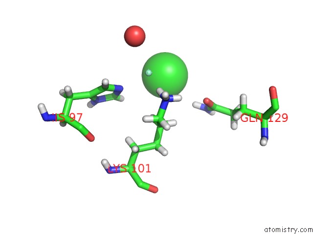
Mono view
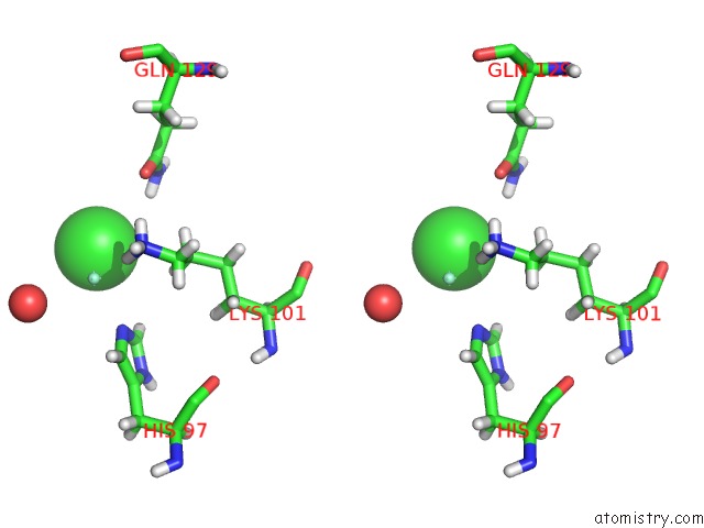
Stereo pair view
|
A full contact list of Chlorine with other atoms in the Cl binding
site number 2 of The Structure of the L-Tryptophan Oxidase Vioa From Chromobacterium Violaceum - Samarium Derivative within 5.0Å range:
probe
|
atom
|
residue
|
distance (Å)
|
B
|
Occ
|
A:Cl437
b:42.1
occ:1.00
|
SM
|
A:SM420
|
2.7
|
35.0
|
1.0
|
HE22
|
A:GLN129
|
3.2
|
41.9
|
1.0
|
HZ1
|
A:LYS101
|
3.7
|
29.5
|
1.0
|
HE1
|
A:HIS97
|
3.7
|
26.8
|
1.0
|
HD3
|
A:LYS101
|
3.7
|
29.3
|
1.0
|
NE2
|
A:HIS97
|
4.0
|
21.2
|
1.0
|
NE2
|
A:GLN129
|
4.0
|
34.9
|
1.0
|
HD2
|
A:LYS101
|
4.1
|
29.3
|
1.0
|
CE1
|
A:HIS97
|
4.2
|
22.4
|
1.0
|
HZ3
|
A:LYS101
|
4.2
|
29.5
|
1.0
|
NZ
|
A:LYS101
|
4.3
|
24.6
|
1.0
|
OE1
|
A:GLN129
|
4.3
|
35.0
|
1.0
|
CD
|
A:LYS101
|
4.4
|
24.4
|
1.0
|
O
|
A:HOH2181
|
4.5
|
31.1
|
1.0
|
HE21
|
A:GLN129
|
4.6
|
41.9
|
1.0
|
CD
|
A:GLN129
|
4.6
|
34.4
|
1.0
|
CE
|
A:LYS101
|
5.0
|
24.1
|
1.0
|
|
Chlorine binding site 3 out
of 3 in 5g3s
Go back to
Chlorine Binding Sites List in 5g3s
Chlorine binding site 3 out
of 3 in the The Structure of the L-Tryptophan Oxidase Vioa From Chromobacterium Violaceum - Samarium Derivative
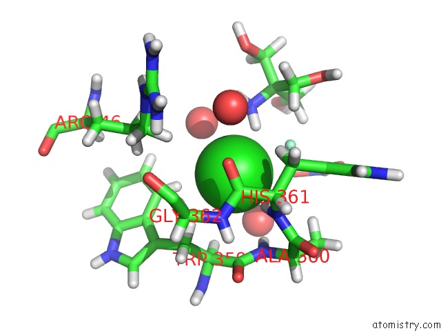
Mono view
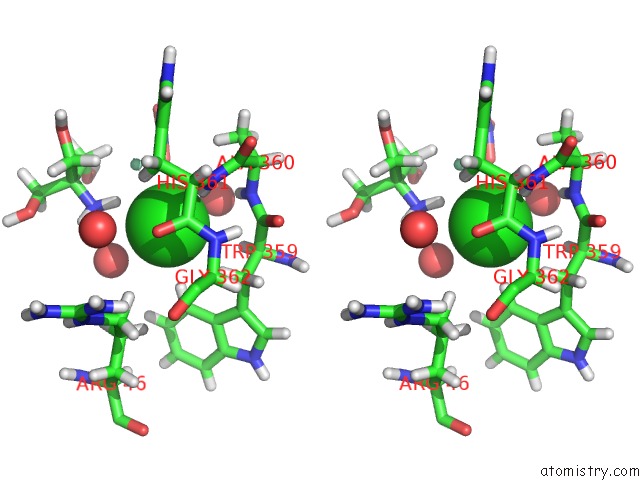
Stereo pair view
|
A full contact list of Chlorine with other atoms in the Cl binding
site number 3 of The Structure of the L-Tryptophan Oxidase Vioa From Chromobacterium Violaceum - Samarium Derivative within 5.0Å range:
probe
|
atom
|
residue
|
distance (Å)
|
B
|
Occ
|
B:Cl432
b:23.3
occ:1.00
|
H
|
B:HIS361
|
2.3
|
26.7
|
1.0
|
HN2
|
B:TRS433
|
2.5
|
45.5
|
1.0
|
SM
|
B:SM420
|
2.9
|
35.2
|
1.0
|
HB3
|
B:TRP359
|
3.0
|
19.9
|
1.0
|
HE3
|
B:TRP359
|
3.0
|
20.2
|
1.0
|
N
|
B:HIS361
|
3.1
|
22.2
|
1.0
|
HB2
|
B:HIS361
|
3.2
|
27.2
|
1.0
|
O
|
B:HOH2029
|
3.3
|
38.5
|
1.0
|
N
|
B:TRS433
|
3.4
|
37.9
|
1.0
|
O
|
B:HOH2195
|
3.5
|
42.0
|
1.0
|
C
|
B:HIS361
|
3.5
|
21.9
|
1.0
|
H
|
B:ALA360
|
3.5
|
22.8
|
1.0
|
HN3
|
B:TRS433
|
3.6
|
45.5
|
1.0
|
HA
|
B:TRP359
|
3.6
|
22.0
|
1.0
|
O
|
A:HOH2123
|
3.6
|
23.9
|
1.0
|
O
|
B:HIS361
|
3.6
|
22.1
|
1.0
|
CA
|
B:HIS361
|
3.6
|
22.7
|
1.0
|
HN1
|
B:TRS433
|
3.7
|
45.5
|
1.0
|
N
|
B:ALA360
|
3.7
|
19.0
|
1.0
|
HA2
|
B:GLY362
|
3.8
|
24.1
|
1.0
|
CB
|
B:HIS361
|
3.8
|
22.6
|
1.0
|
CB
|
B:TRP359
|
3.9
|
16.6
|
1.0
|
HD3
|
B:ARG46
|
3.9
|
24.2
|
1.0
|
O3
|
B:NO3431
|
3.9
|
29.5
|
1.0
|
N
|
B:GLY362
|
3.9
|
21.6
|
1.0
|
C
|
B:TRP359
|
3.9
|
20.2
|
1.0
|
CE3
|
B:TRP359
|
4.0
|
16.9
|
1.0
|
CA
|
B:TRP359
|
4.0
|
18.3
|
1.0
|
H32
|
B:TRS433
|
4.0
|
45.5
|
1.0
|
HB1
|
B:ALA360
|
4.2
|
24.1
|
1.0
|
C
|
B:ALA360
|
4.2
|
20.4
|
1.0
|
H
|
B:GLY362
|
4.3
|
25.9
|
1.0
|
ND1
|
B:HIS361
|
4.4
|
24.8
|
1.0
|
CA
|
B:GLY362
|
4.4
|
20.0
|
1.0
|
HG2
|
B:ARG46
|
4.4
|
24.4
|
1.0
|
CA
|
B:ALA360
|
4.4
|
19.6
|
1.0
|
HB2
|
B:TRP359
|
4.5
|
19.9
|
1.0
|
CG
|
B:HIS361
|
4.6
|
24.2
|
1.0
|
HA
|
B:HIS361
|
4.6
|
27.3
|
1.0
|
HB3
|
B:HIS361
|
4.6
|
27.2
|
1.0
|
C
|
B:TRS433
|
4.6
|
35.9
|
1.0
|
O
|
B:TRP359
|
4.7
|
21.6
|
1.0
|
CD2
|
B:TRP359
|
4.7
|
15.6
|
1.0
|
H22
|
B:TRS433
|
4.7
|
43.7
|
1.0
|
CG
|
B:TRP359
|
4.7
|
15.5
|
1.0
|
CD
|
B:ARG46
|
4.7
|
20.2
|
1.0
|
HG3
|
B:ARG46
|
4.8
|
24.4
|
1.0
|
C3
|
B:TRS433
|
4.8
|
37.9
|
1.0
|
CB
|
B:ALA360
|
4.9
|
20.1
|
1.0
|
O2
|
B:NO3430
|
4.9
|
53.0
|
1.0
|
HZ3
|
B:TRP359
|
4.9
|
20.9
|
1.0
|
CG
|
B:ARG46
|
4.9
|
20.3
|
1.0
|
CZ3
|
B:TRP359
|
4.9
|
17.4
|
1.0
|
HA3
|
B:GLY362
|
5.0
|
24.1
|
1.0
|
|
Reference:
J.Fuller,
R.Roepke,
J.Krausze,
K.E.Rennhack,
N.P.Daniel,
W.Blankenfeldt,
S.Schulz,
D.Jahn,
J.Moser.
Biosynthesis of Violacein: Structure and Function of L-Tryptophan Oxidase Vioa Chromobacterium Violaceum J.Biol.Chem. V. 291 20068 2016.
ISSN: ISSN 0021-9258
PubMed: 27466367
DOI: 10.1074/JBC.M116.741561
Page generated: Fri Jul 26 08:19:28 2024
|