Chlorine »
PDB 5mzf-5n6t »
5n5r »
Chlorine in PDB 5n5r: 14-3-3 Sigma in Complex with Taz PS89 Peptide and Fragment NV1
Protein crystallography data
The structure of 14-3-3 Sigma in Complex with Taz PS89 Peptide and Fragment NV1, PDB code: 5n5r
was solved by
E.Sijbesma,
S.Leysen,
C.Ottmann,
with X-Ray Crystallography technique. A brief refinement statistics is given in the table below:
| Resolution Low / High (Å) | 41.74 / 1.80 |
| Space group | C 2 2 21 |
| Cell size a, b, c (Å), α, β, γ (°) | 81.962, 111.974, 62.630, 90.00, 90.00, 90.00 |
| R / Rfree (%) | 14.9 / 18.2 |
Other elements in 5n5r:
The structure of 14-3-3 Sigma in Complex with Taz PS89 Peptide and Fragment NV1 also contains other interesting chemical elements:
| Magnesium | (Mg) | 3 atoms |
Chlorine Binding Sites:
The binding sites of Chlorine atom in the 14-3-3 Sigma in Complex with Taz PS89 Peptide and Fragment NV1
(pdb code 5n5r). This binding sites where shown within
5.0 Angstroms radius around Chlorine atom.
In total only one binding site of Chlorine was determined in the 14-3-3 Sigma in Complex with Taz PS89 Peptide and Fragment NV1, PDB code: 5n5r:
In total only one binding site of Chlorine was determined in the 14-3-3 Sigma in Complex with Taz PS89 Peptide and Fragment NV1, PDB code: 5n5r:
Chlorine binding site 1 out of 1 in 5n5r
Go back to
Chlorine binding site 1 out
of 1 in the 14-3-3 Sigma in Complex with Taz PS89 Peptide and Fragment NV1
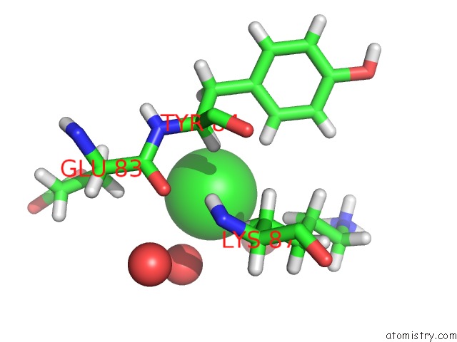
Mono view
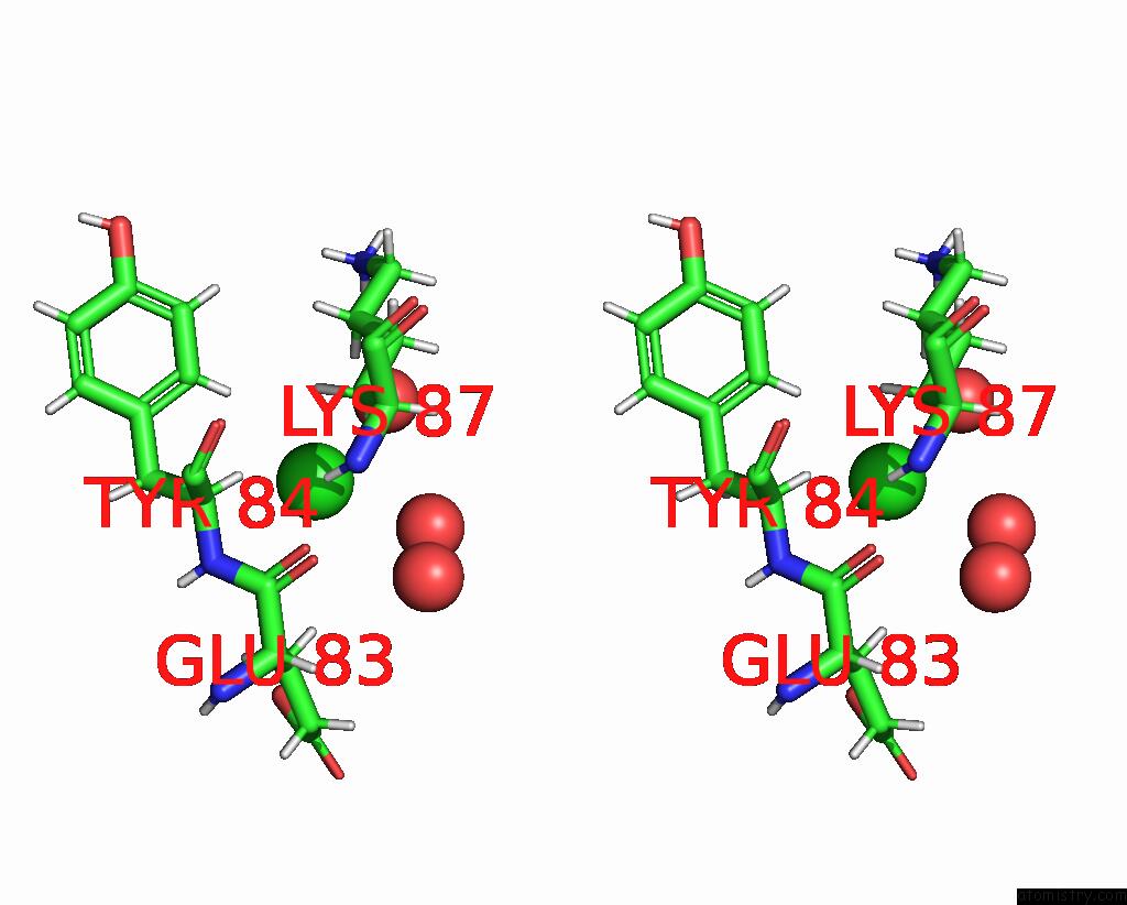
Stereo pair view
Mono view
Stereo pair view
A full contact list of Chlorine with other atoms in the Cl binding
site number 1 of 14-3-3 Sigma in Complex with Taz PS89 Peptide and Fragment NV1 within 5.0Å range:
|
Reference:
E.Sijbesma,
L.Skora,
S.Leysen,
L.Brunsveld,
U.Koch,
P.Nussbaumer,
W.Jahnke,
C.Ottmann.
Identification of Two Secondary Ligand Binding Sites in 14-3-3 Proteins Using Fragment Screening. Biochemistry V. 56 3972 2017.
ISSN: ISSN 1520-4995
PubMed: 28681606
DOI: 10.1021/ACS.BIOCHEM.7B00153
Page generated: Sat Jul 12 05:52:33 2025
ISSN: ISSN 1520-4995
PubMed: 28681606
DOI: 10.1021/ACS.BIOCHEM.7B00153
Last articles
Cl in 7ZKSCl in 7ZKX
Cl in 7ZKR
Cl in 7ZJU
Cl in 7ZIV
Cl in 7ZJT
Cl in 7ZIK
Cl in 7ZIZ
Cl in 7ZI0
Cl in 7ZIC